Deposition Date
1997-04-30
Release Date
1998-03-18
Last Version Date
2024-04-03
Entry Detail
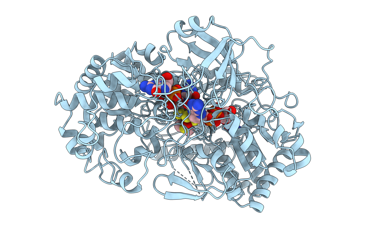
PDB ID:
4DMR
Keywords:
Title:
REDUCED DMSO REDUCTASE FROM RHODOBACTER CAPSULATUS WITH BOUND DMSO SUBSTRATE
Biological Source:
Source Organism(s):
Rhodobacter capsulatus (Taxon ID: 1061)
Method Details:
Experimental Method:
Resolution:
1.90 Å
R-Value Free:
0.19
R-Value Work:
0.15
Space Group:
P 41 21 2