4DL1 image
Deposition Date 2012-02-05
Release Date 2012-03-21
Last Version Date 2024-12-25
Entry Detail
PDB ID:
4DL1
Keywords:
Title:
Crystal Structure of human Myeloperoxidase with covalent thioxanthine analog
Biological Source:
Source Organism:
Homo sapiens (Taxon ID: 9606)
Method Details:
Experimental Method:
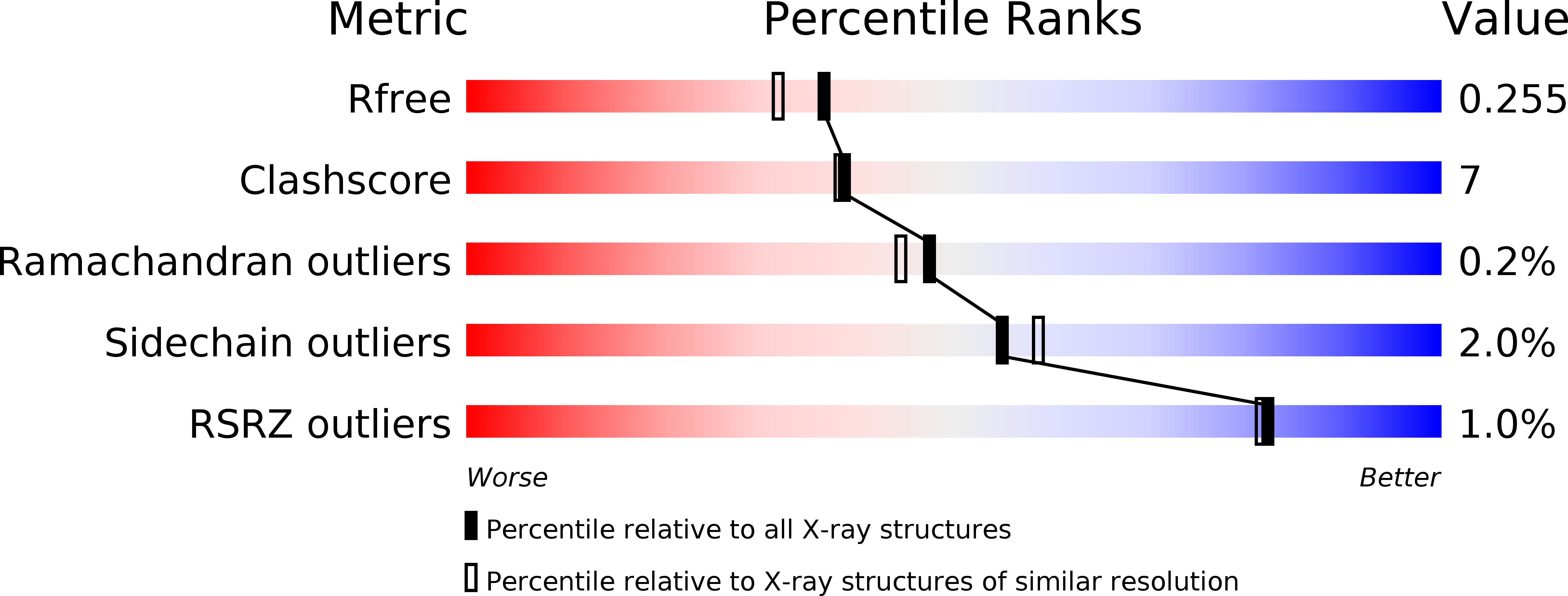
Resolution:
2.00 Å
R-Value Free:
0.24
R-Value Work:
0.19
R-Value Observed:
0.19
Space Group:
P 1 21 1
Macromolecular Entities
Structures with similar UniProt ID
Protein Blast
Polymer Type:polypeptide(L)
Molecule:Myeloperoxidase light chain
Gene (Uniprot):MPO
Chain IDs:A, C (auth: B), E, G (auth: F), I, K (auth: J), M, O (auth: N)
Chain Length:104
Number of Molecules:8
Biological Source:Homo sapiens
Structures with similar UniProt ID
Protein Blast
Polymer Type:polypeptide(L)
Molecule:Myeloperoxidase heavy chain
Gene (Uniprot):MPO
Chain IDs:B (auth: C), D, F (auth: G), H, J (auth: K), L, N (auth: O), P
Chain Length:466
Number of Molecules:8
Biological Source:Homo sapiens
Modified Residue
Compound ID Chain ID Parent Comp ID Details 2D Image
ASN B ASN GLYCOSYLATION SITE
CSO B CYS S-HYDROXYCYSTEINE
Primary Citation
Deconstruction of activity-dependent covalent modification of heme in human neutrophil myeloperoxidase by multistage mass spectrometry (MS(4)).
Biochemistry 51 2065 2077 (2012)
PMID: 22352991 DOI: 10.1021/bi201872j

Abstact

Myeloperoxidase (MPO) is known to be inactivated and covalently modified by treatment with hydrogen peroxide and agents similar to 3-(2-ethoxypropyl)-2-thioxo-2,3-dihydro-1H-purin-6(9H)-one (1), a 254.08 Da derivative of 2-thioxanthine. Peptide mapping by liquid chromatography and mass spectrometry detected modification by 1 in a labile peptide-heme-peptide fragment of the enzyme, accompanied by a mass increase of 252.08 Da. The loss of two hydrogen atoms was consistent with mechanism-based oxidative coupling. Multistage mass spectrometry (MS(4)) of the modified fragment in an ion trap/Orbitrap spectrometer demonstrated that 1 was coupled directly to heme. Use of a 10 amu window delivered the full isotopic envelope of each precursor ion to collision-induced dissociation, preserving definitive isotopic profiles for iron-containing fragments through successive steps of multistage mass spectrometry. Iron isotope signatures and accurate mass measurements supported the structural assignments. Crystallographic analysis confirmed linkage between the methyl substituent of the heme pyrrole D ring and the sulfur atom of 1. The final orientation of 1 perpendicular to the plane of the heme ring suggested a mechanism consisting of two consecutive one-electron oxidations of 1 by MPO. Multistage mass spectrometry using stage-specific collision energies permits stepwise deconstruction of modifications of heme enzymes containing covalent links between the heme group and the polypeptide chain.

Legend

Protein

Chemical

Disease

Primary Citation of related structures
Feedback Form
Name
Email
Institute
Feedback