Deposition Date
2012-02-03
Release Date
2012-03-21
Last Version Date
2024-11-27
Entry Detail
PDB ID:
4DKL
Keywords:
Title:
Crystal structure of the mu-opioid receptor bound to a morphinan antagonist
Biological Source:
Source Organism(s):
Mus musculus (Taxon ID: 10090)
Enterobacteria phage T4 (Taxon ID: 10665)
Enterobacteria phage T4 (Taxon ID: 10665)
Expression System(s):
Method Details:
Experimental Method:
Resolution:
2.80 Å
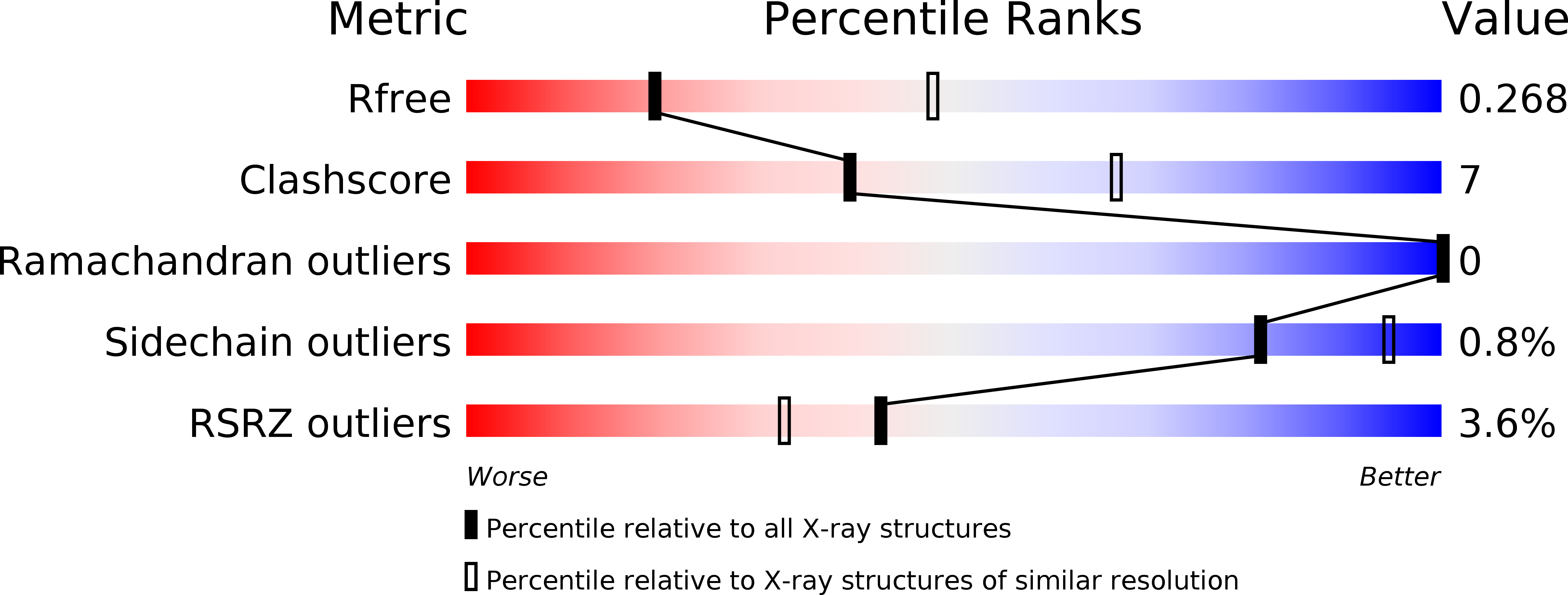
R-Value Free:
0.27
R-Value Work:
0.23
R-Value Observed:
0.23
Space Group:
C 1 2 1