Deposition Date
2012-02-03
Release Date
2013-03-20
Last Version Date
2024-03-13
Entry Detail
PDB ID:
4DKK
Keywords:
Title:
The X-ray Crystal Structure of the Human STAU1 SSM-'RBD'5 Domain-Swapped Dimer
Biological Source:
Source Organism(s):
Homo sapiens (Taxon ID: 9606)
Expression System(s):
Method Details:
Experimental Method:
Resolution:
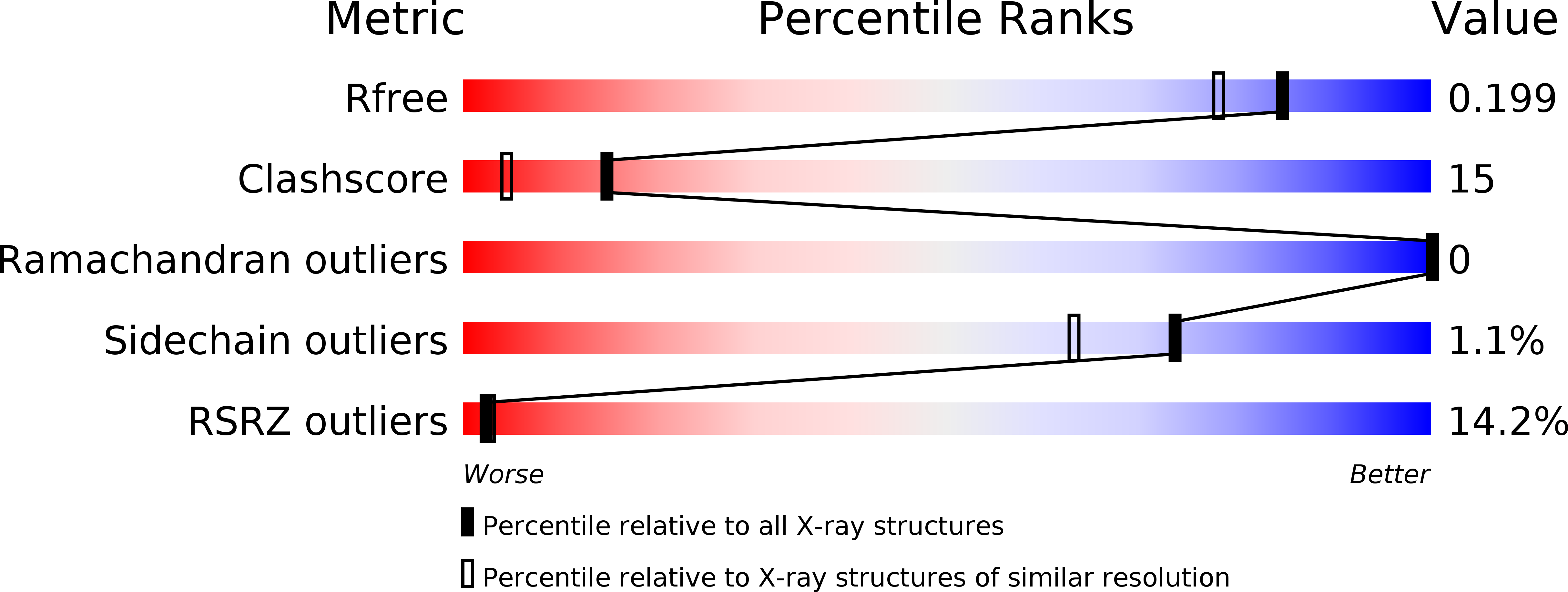
1.70 Å
R-Value Free:
0.20
R-Value Work:
0.16
R-Value Observed:
0.16
Space Group:
P 41 21 2