Deposition Date
2012-02-03
Release Date
2012-03-21
Last Version Date
2024-10-30
Entry Detail
PDB ID:
4DK7
Keywords:
Title:
Crystal structure of LXR ligand binding domain in complex with full agonist 1
Biological Source:
Source Organism(s):
Homo sapiens (Taxon ID: 9606)
Expression System(s):
Method Details:
Experimental Method:
Resolution:
2.45 Å
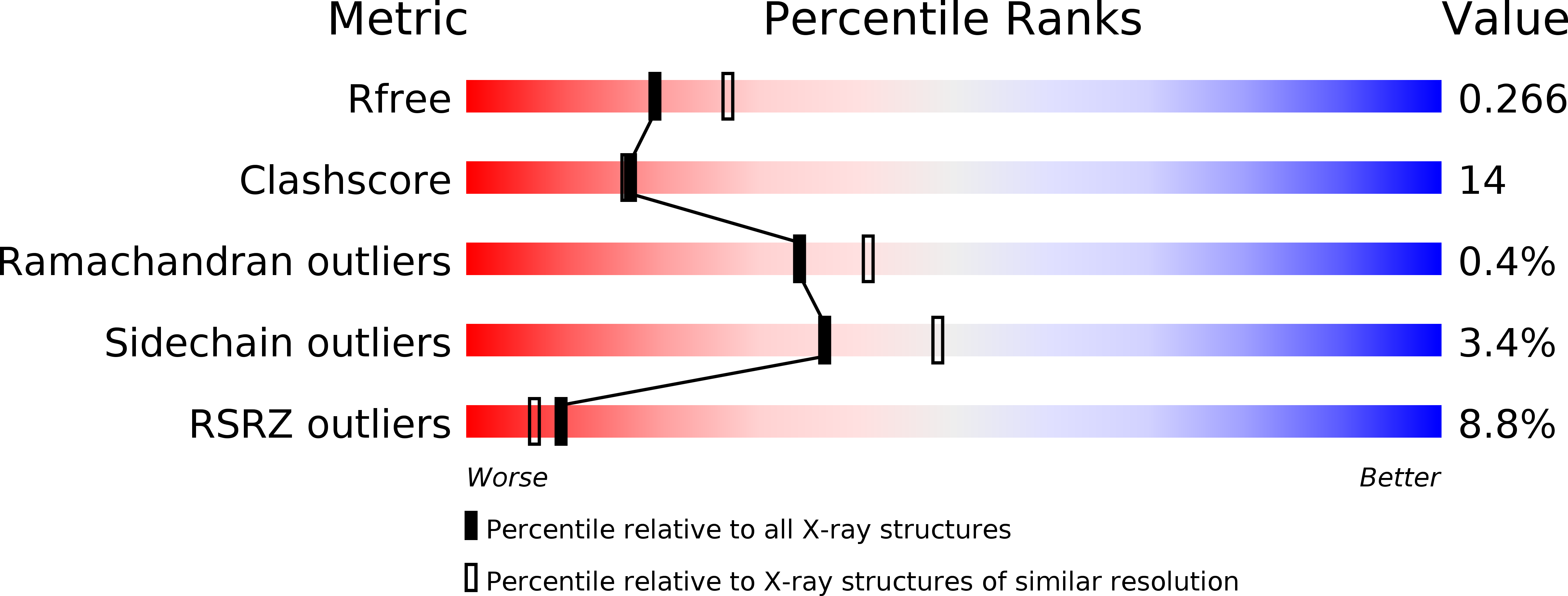
R-Value Free:
0.27
R-Value Work:
0.23
R-Value Observed:
0.27
Space Group:
P 43 21 2