Deposition Date
2012-01-30
Release Date
2012-02-08
Last Version Date
2023-09-13
Entry Detail
PDB ID:
4DHY
Keywords:
Title:
Crystal structure of human glucokinase in complex with glucose and activator
Biological Source:
Source Organism(s):
Homo sapiens (Taxon ID: 9606)
Expression System(s):
Method Details:
Experimental Method:
Resolution:
2.38 Å
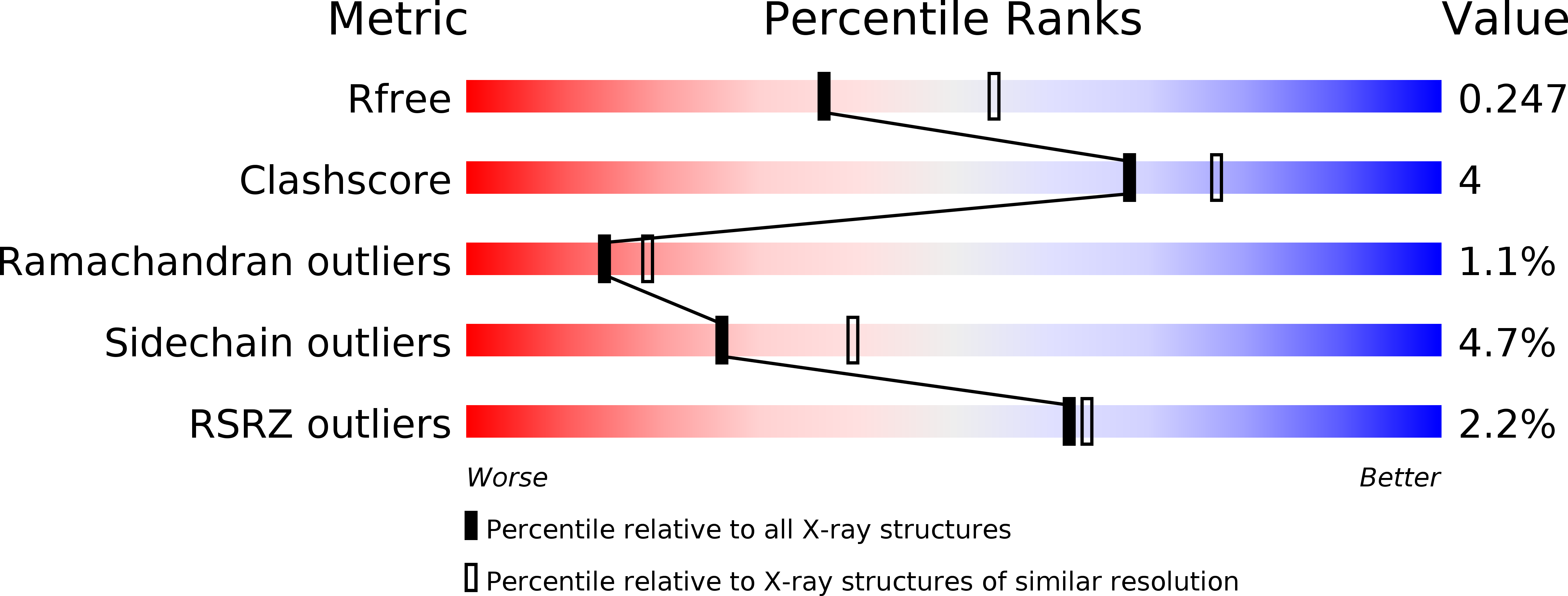
R-Value Free:
0.24
R-Value Work:
0.18
R-Value Observed:
0.18
Space Group:
P 21 21 21