Deposition Date
2012-01-23
Release Date
2012-03-21
Last Version Date
2023-09-13
Entry Detail
PDB ID:
4DFG
Keywords:
Title:
Crystal Structure of Wild-type HIV-1 Protease with Cyclopentyltetrahydro- furanyl Urethanes as P2-ligand, GRL-0249A
Biological Source:
Source Organism(s):
Human immunodeficiency virus type 1 (Taxon ID: 11686)
Expression System(s):
Method Details:
Experimental Method:
Resolution:
1.23 Å
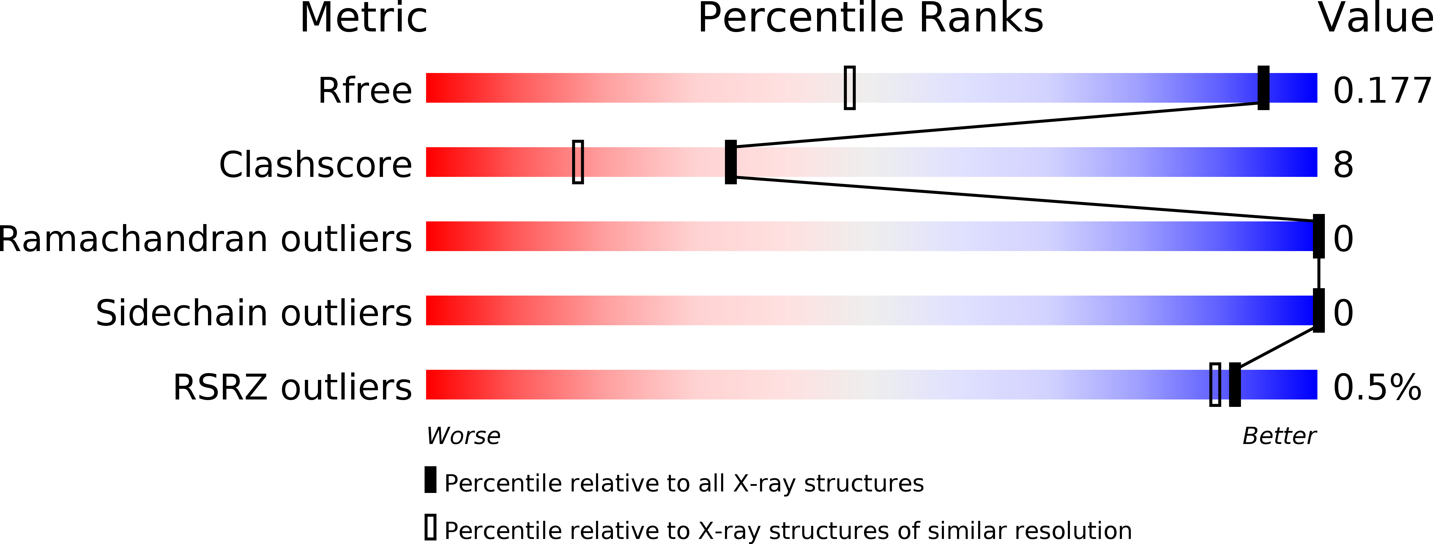
R-Value Free:
0.18
R-Value Work:
0.14
R-Value Observed:
0.14
Space Group:
P 21 21 2