Planned Maintenance: Some services may turn out to be unavailable from 15th January, 2026 to 16th January, 2026. We apologize for the inconvenience!

4DF3 image
Deposition Date 2012-01-22
Release Date 2012-08-08
Last Version Date 2024-02-28
Entry Detail
PDB ID:
4DF3
Keywords:
Title:
Crystal Structure of Aeropyrum pernix fibrillarin in complex with natively bound S-adenosyl-L-methionine at 1.7A
Method Details:
Experimental Method:
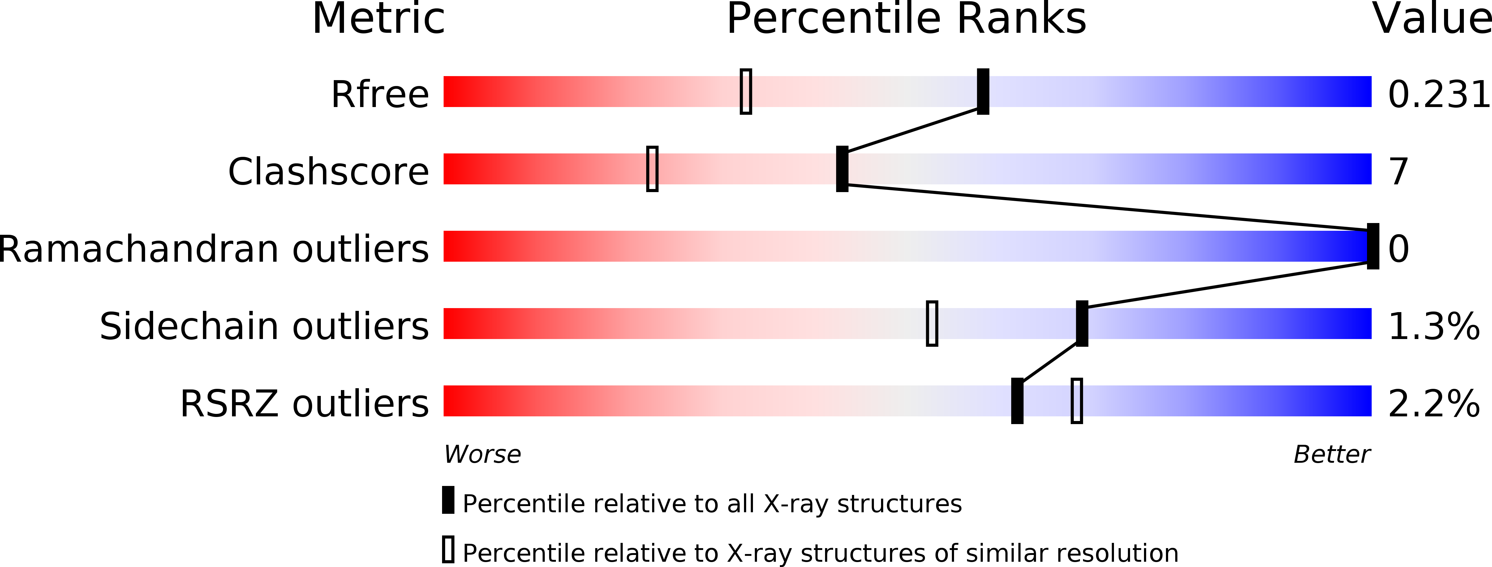
Resolution:
1.73 Å
R-Value Free:
0.23
R-Value Work:
0.18
R-Value Observed:
0.18
Space Group:
P 1
Macromolecular Entities
Structures with similar UniProt ID
Protein Blast
Polymer Type:polypeptide(L)
Molecule:Fibrillarin-like rRNA/tRNA 2'-O-methyltransferase
Gene (Uniprot):flpA
Mutagens:R8S, E9R
Chain IDs:A, B
Chain Length:233
Number of Molecules:2
Biological Source:Aeropyrum pernix
Ligand Molecules
Primary Citation
Structure of Aeropyrum pernix fibrillarin in complex with natively bound S-adenosyl-L-methionine at 1.7 A resolution.
Acta Crystallogr.,Sect.F 68 854 859 (2012)
PMID: 22869109 DOI: 10.1107/S1744309112026528

Abstact

Fibrillarin is the key methyltransferase associated with the C/D class of small nuclear ribonucleoproteins (snRNPs) and participates in the preliminary step of pre-ribosomal rRNA processing. This molecule is found in the fibrillar regions of the eukaryotic nucleolus and is involved in methylation of the 2'-O atom of ribose in rRNA. Human fibrillarin contains an N-terminal GAR domain, a central RNA-binding domain comprising an RNP-2-like superfamily consensus sequence and a catalytic C-terminal helical domain. Here, Aeropyrum pernix fibrillarin is described, which is homologous to the C-terminal domain of human fibrillarin. The protein was crystallized with an S-adenosyl-L-methionine (SAM) ligand bound in the active site. The molecular structure of this complex was solved using X-ray crystallography at a resolution of 1.7 Å using molecular replacement with fibrillarin structural homologs. The structure shows the atomic details of SAM and its active-site interactions; there are a number of conserved residues that interact directly with the cofactor. Notably, the adenine ring of SAM is stabilized by π-π interactions with the conserved residue Phe110 and by electrostatic interactions with the Asp134, Ala135 and Gln157 residues. The π-π interaction appears to play a critical role in stabilizing the association of SAM with fibrillarin. Furthermore, comparison of A. pernix fibrillarin with homologous structures revealed different orientations of Phe110 and changes in α-helix 6 of fibrillarin and suggests key differences in its interactions with the adenine ring of SAM in the active site and with the C/D RNA. These differences may play a key role in orienting the SAM ligand for catalysis as well as in the assembly of other ribonucleoproteins and in the interactions with C/D RNA.

Legend

Protein

Chemical

Disease

Primary Citation of related structures
Feedback Form
Name
Email
Institute
Feedback