4DDZ image
Entry Detail
PDB ID:
4DDZ
Keywords:
Title:
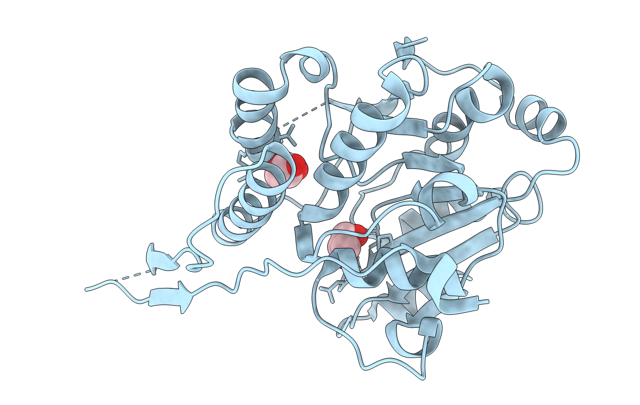
Crystal structure of glucosyl-3-phosphoglycerate synthase from Mycobacterium tuberculosis
Biological Source:
Host Organism:
PDB Version:
Deposition Date:
2012-01-19
Release Date:
2012-06-06
Method Details:
Experimental Method:
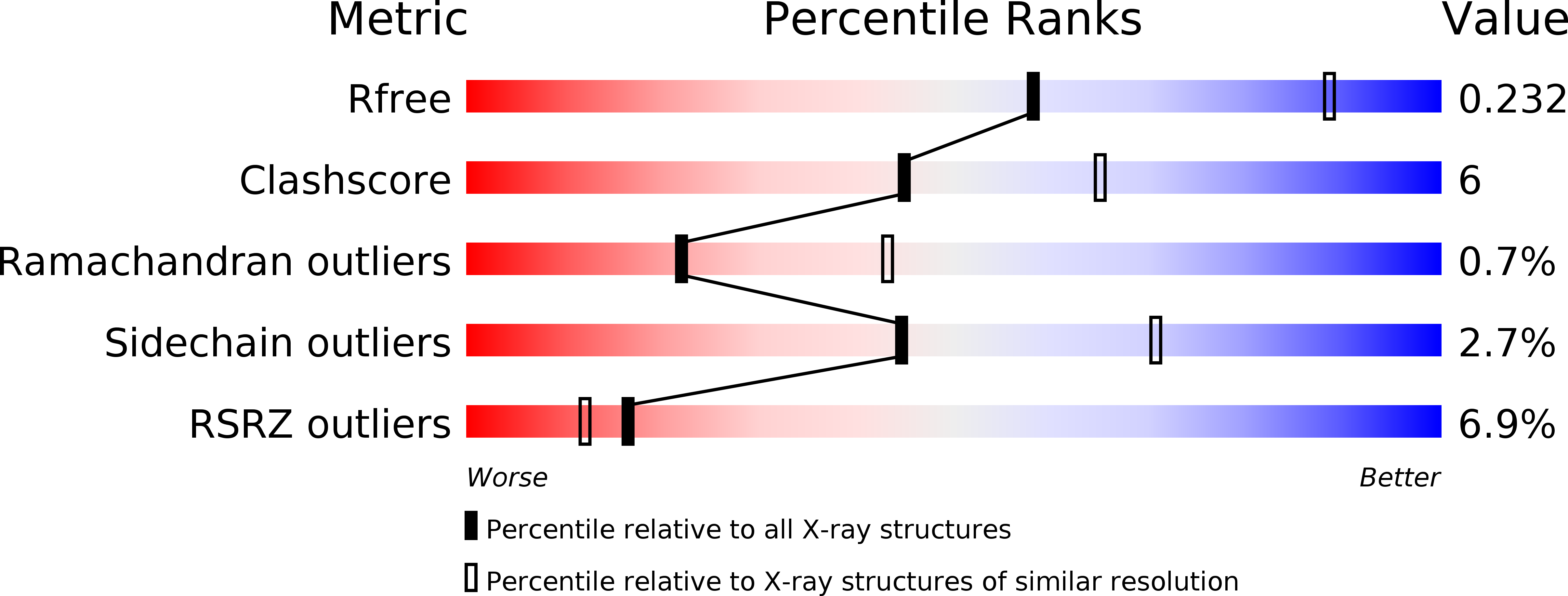
Resolution:
2.60 Å
R-Value Free:
0.24
R-Value Work:
0.21
R-Value Observed:
0.21
Space Group:
I 41
Macromolecular Entities
Polymer Type:polypeptide(L)
Description:GLUCOSYL-3-PHOSPHOGLYCERATE SYNTHASE (GpgS)
Chain IDs:A
Chain Length:344
Number of Molecules:1
Biological Source:Mycobacterium tuberculosis
Ligand Molecules
Primary Citation
Mechanistic insights into the retaining glucosyl-3-phosphoglycerate synthase from mycobacteria.
J.Biol.Chem. 287 24649 24661 (2012)
PMID: 22637481 DOI: 10.1074/jbc.M112.368191

Abstact

Considerable progress has been made in recent years in our understanding of the structural basis of glycosyl transfer. Yet the nature and relevance of the conformational changes associated with substrate recognition and catalysis remain poorly understood. We have focused on the glucosyl-3-phosphoglycerate synthase (GpgS), a "retaining" enzyme, that initiates the biosynthetic pathway of methylglucose lipopolysaccharides in mycobacteria. Evidence is provided that GpgS displays an unusually broad metal ion specificity for a GT-A enzyme, with Mg(2+), Mn(2+), Ca(2+), Co(2+), and Fe(2+) assisting catalysis. In the crystal structure of the apo-form of GpgS, we have observed that a flexible loop adopts a double conformation L(A) and L(I) in the active site of both monomers of the protein dimer. Notably, the L(A) loop geometry corresponds to an active conformation and is conserved in two other relevant states of the enzyme, namely the GpgS·metal·nucleotide sugar donor and the GpgS·metal·nucleotide·acceptor-bound complexes, indicating that GpgS is intrinsically in a catalytically active conformation. The crystal structure of GpgS in the presence of Mn(2+)·UDP·phosphoglyceric acid revealed an alternate conformation for the nucleotide sugar β-phosphate, which likely occurs upon sugar transfer. Structural, biochemical, and biophysical data point to a crucial role of the β-phosphate in donor and acceptor substrate binding and catalysis. Altogether, our experimental data suggest a model wherein the catalytic site is essentially preformed, with a few conformational changes of lateral chain residues as the protein proceeds along the catalytic cycle. This model of action may be applicable to a broad range of GT-A glycosyltransferases.

Legend

Protein

Chemical

Disease

Primary Citation of related structures