Deposition Date
2012-01-17
Release Date
2012-12-12
Last Version Date
2025-03-26
Entry Detail
PDB ID:
4DCJ
Keywords:
Title:
Crystal structure of caspase 3, L168D mutant
Biological Source:
Source Organism(s):
Homo sapiens (Taxon ID: 9606)
Expression System(s):
Method Details:
Experimental Method:
Resolution:
1.70 Å
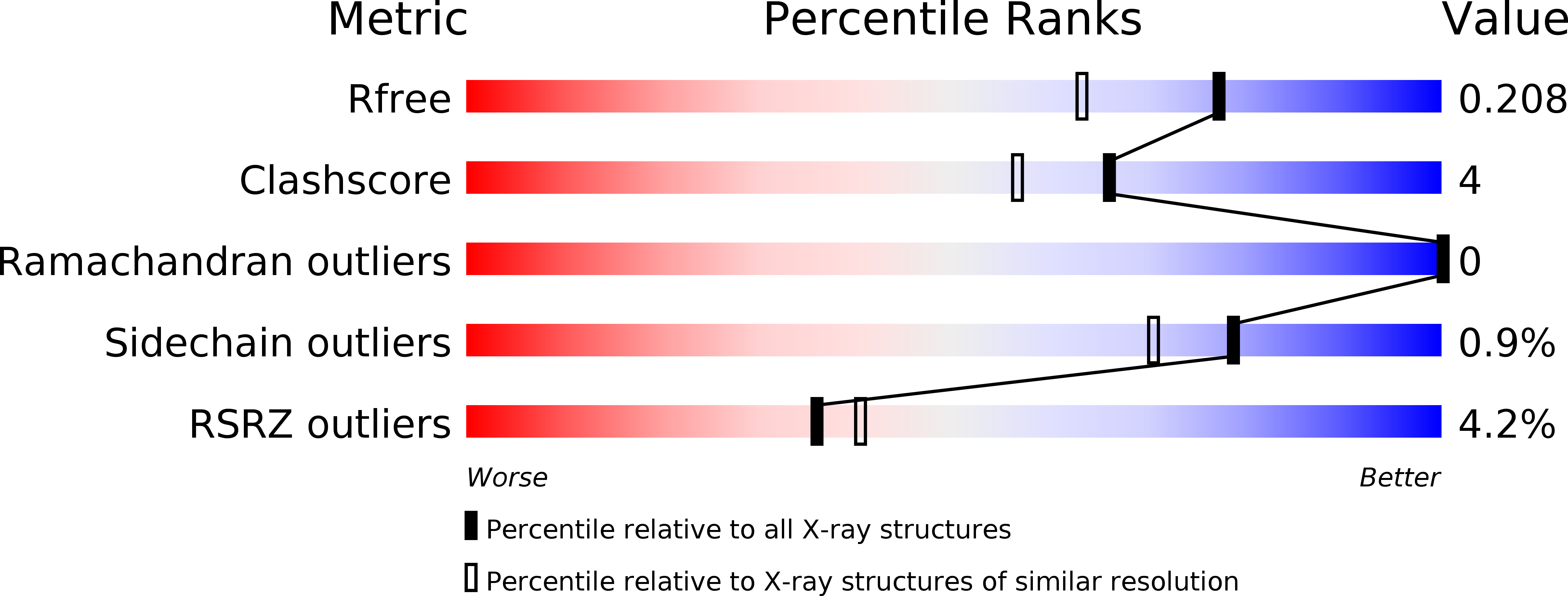
R-Value Free:
0.21
R-Value Work:
0.19
R-Value Observed:
0.19
Space Group:
P 1 21 1