Deposition Date
2012-01-17
Release Date
2013-01-23
Last Version Date
2024-10-09
Entry Detail
PDB ID:
4DC5
Keywords:
Title:
Crystal Structure of Thaumatin Unexposed to Excessive SONICC Imaging Laser Dose.
Biological Source:
Source Organism(s):
Thaumatococcus daniellii (Taxon ID: 4621)
Method Details:
Experimental Method:
Resolution:
1.48 Å
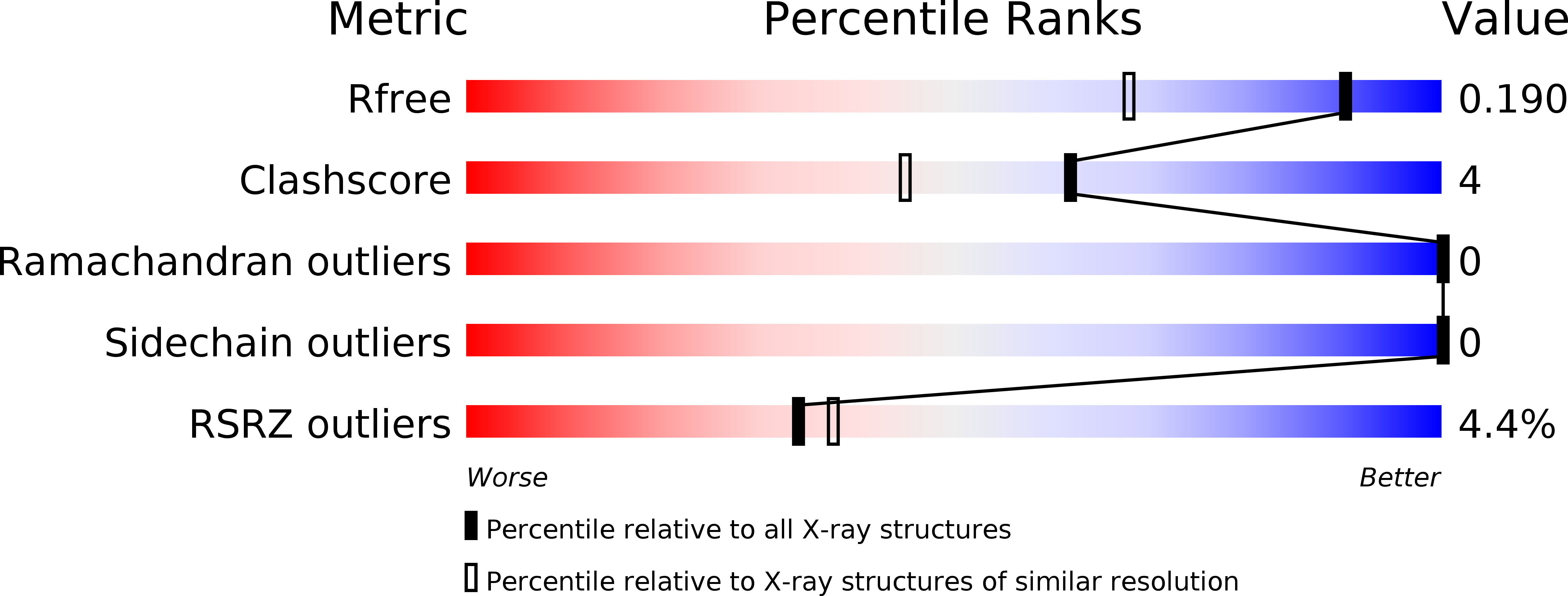
R-Value Free:
0.19
R-Value Work:
0.17
R-Value Observed:
0.17
Space Group:
P 41 21 2