4DAT image
Deposition Date 2012-01-13
Release Date 2012-06-13
Last Version Date 2024-10-30
Entry Detail
PDB ID:
4DAT
Title:
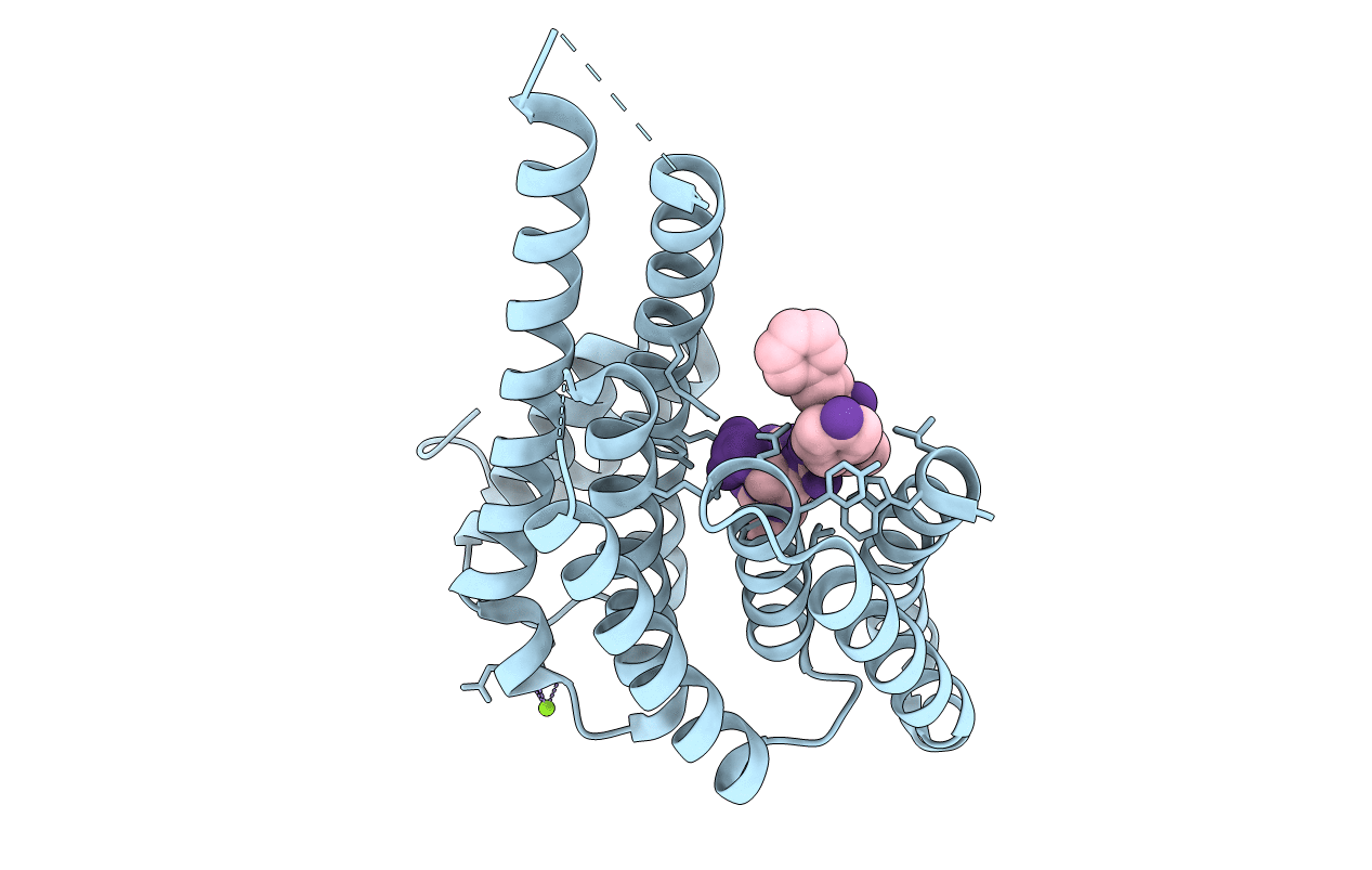
Structure of 14-3-3 sigma in complex with PADI6 14-3-3 binding motif II
Biological Source:
Source Organism:
Homo sapiens (Taxon ID: 9606)
Host Organism:
Method Details:
Experimental Method:
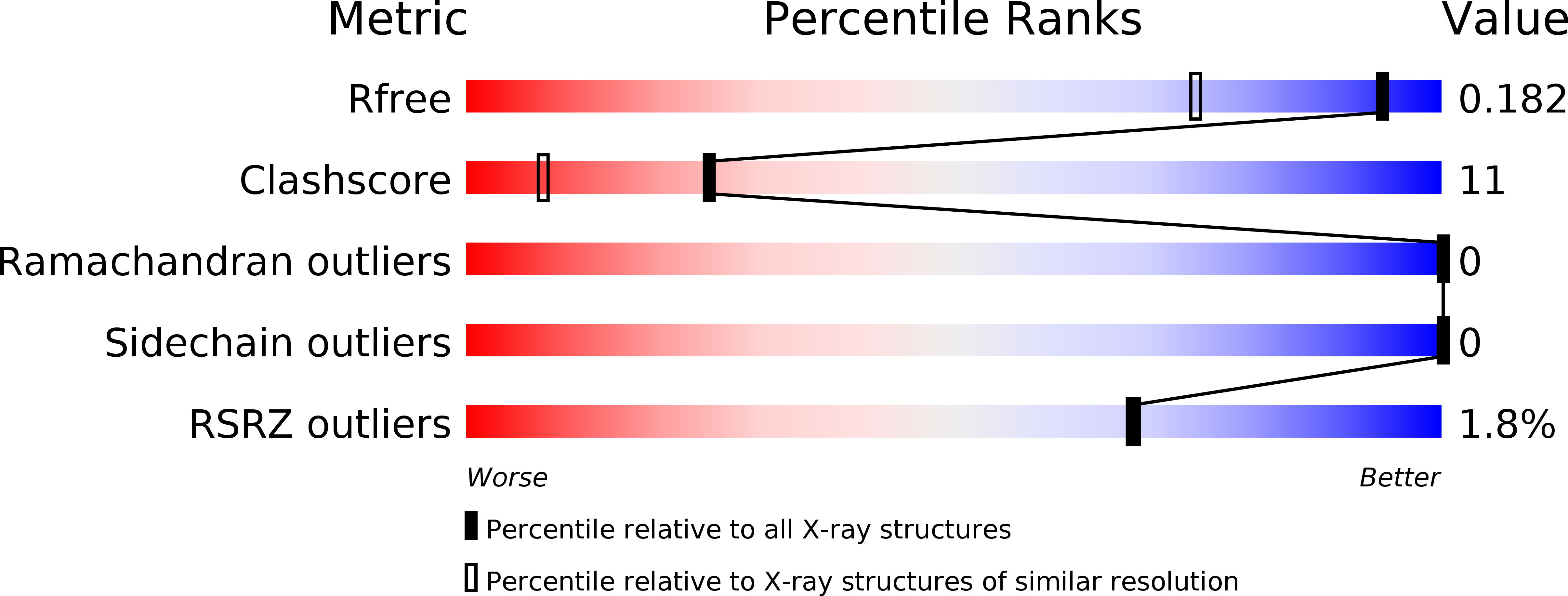
Resolution:
1.40 Å
R-Value Free:
0.18
R-Value Work:
0.14
R-Value Observed:
0.14
Space Group:
C 2 2 21
Macromolecular Entities
Polymer Type:polypeptide(L)
Molecule:14-3-3 protein sigma
Gene (Uniprot):SFN
Chain IDs:A
Chain Length:234
Number of Molecules:1
Biological Source:Homo sapiens
Polymer Type:polypeptide(L)
Molecule:Peptidylarginine Deiminase type VI
Chain IDs:B
Chain Length:9
Number of Molecules:1
Biological Source:Homo sapiens
Modified Residue
Compound ID Chain ID Parent Comp ID Details 2D Image
SEP B SER PHOSPHOSERINE
Ligand Molecules
Primary Citation
Identification and structural characterization of two 14-3-3 binding sites in the human peptidylarginine deiminase type VI.
J.Struct.Biol. 180 65 72 (2012)
PMID: 22634725 DOI: 10.1016/j.jsb.2012.05.010

Abstact

The regulation and function of peptidylarginine deiminase isoform VI (PAD6), which is a highly abundant protein associated with the cytoplasmic lattices in mammalian oocytes, is poorly understood so far. It has been shown previously, that 14-3-3 proteins, a class of regulatory adapter proteins ubiquitous in eukaryotes, bind to PAD6 in vivo in a phosphorylation dependent manner. Here we identify possible 14-3-3 binding sites in human PAD6 by in silico methods, looking for conserved, surface exposed serine residues. Two of these sites were confirmed as 14-3-3 binding sites by fluorescence polarization competition and X-ray crystallography. We furthermore suggest a role of RSK-type kinases in the phosphorylation of one of these two binding sites and provide evidence in the form of in vitro kinase assays with p70S6 kinase and RSK1.

Legend

Protein

Chemical

Disease

Primary Citation of related structures