4DAJ image
Deposition Date 2012-01-12
Release Date 2012-02-22
Last Version Date 2024-10-30
Entry Detail
PDB ID:
4DAJ
Title:
Structure of the M3 Muscarinic Acetylcholine Receptor
Biological Source:
Source Organism:
Host Organism:
Method Details:
Experimental Method:
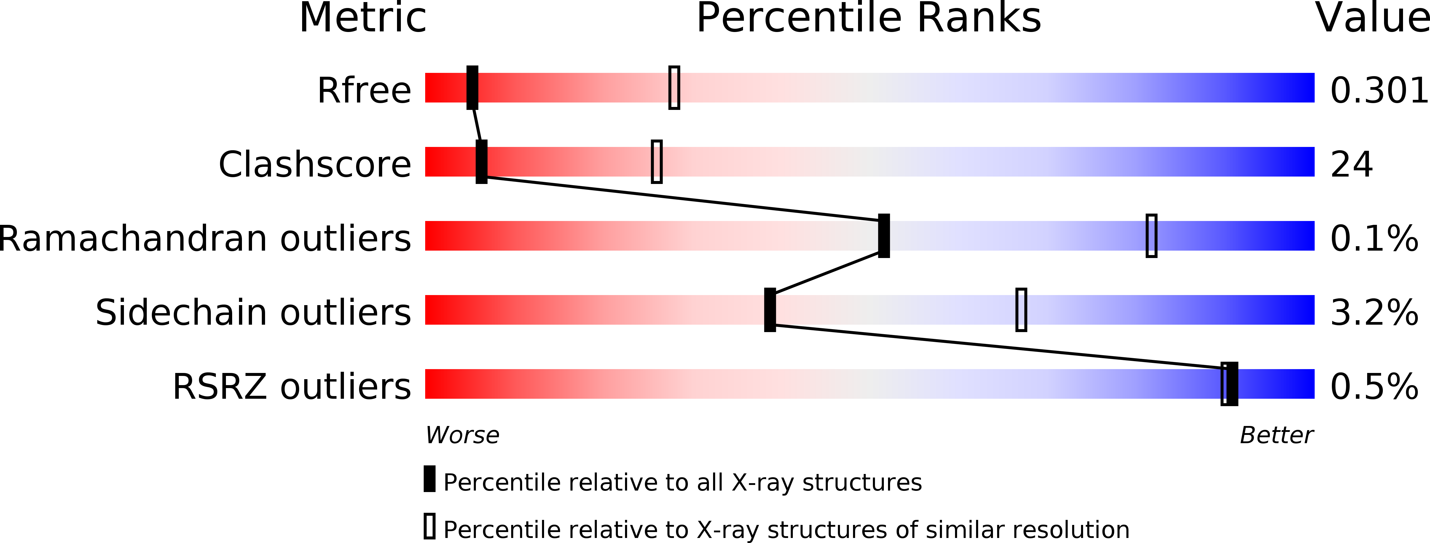
Resolution:
3.40 Å
R-Value Free:
0.30
R-Value Work:
0.25
R-Value Observed:
0.25
Space Group:
P 1
Macromolecular Entities
Protein Blast
Polymer Type:polypeptide(L)
Molecule:Muscarinic acetylcholine receptor M3, Lysozyme
Gene (Uniprot):E, Chrm3
Mutagens:C1054T, C1097A
Chain IDs:A, B, C, D
Chain Length:479
Number of Molecules:4
Biological Source:Rattus norvegicus, Enterobacteria phage T4
Primary Citation
Structure and dynamics of the M3 muscarinic acetylcholine receptor.
Nature 482 552 556 (2012)
PMID: 22358844 DOI: 10.1038/nature10867

Abstact

Acetylcholine, the first neurotransmitter to be identified, exerts many of its physiological actions via activation of a family of G-protein-coupled receptors (GPCRs) known as muscarinic acetylcholine receptors (mAChRs). Although the five mAChR subtypes (M1-M5) share a high degree of sequence homology, they show pronounced differences in G-protein coupling preference and the physiological responses they mediate. Unfortunately, despite decades of effort, no therapeutic agents endowed with clear mAChR subtype selectivity have been developed to exploit these differences. We describe here the structure of the G(q/11)-coupled M3 mAChR ('M3 receptor', from rat) bound to the bronchodilator drug tiotropium and identify the binding mode for this clinically important drug. This structure, together with that of the G(i/o)-coupled M2 receptor, offers possibilities for the design of mAChR subtype-selective ligands. Importantly, the M3 receptor structure allows a structural comparison between two members of a mammalian GPCR subfamily displaying different G-protein coupling selectivities. Furthermore, molecular dynamics simulations suggest that tiotropium binds transiently to an allosteric site en route to the binding pocket of both receptors. These simulations offer a structural view of an allosteric binding mode for an orthosteric GPCR ligand and provide additional opportunities for the design of ligands with different affinities or binding kinetics for different mAChR subtypes. Our findings not only offer insights into the structure and function of one of the most important GPCR families, but may also facilitate the design of improved therapeutics targeting these critical receptors.

Legend

Protein

Chemical

Disease

Primary Citation of related structures