4D8P image
Deposition Date 2012-01-11
Release Date 2012-03-14
Last Version Date 2024-11-20
Entry Detail
PDB ID:
4D8P
Keywords:
Title:
Structural and functional studies of the trans-encoded HLA-DQ2.3 (DQA1*03:01/DQB1*02:01) molecule
Biological Source:
Source Organism:
Homo sapiens (Taxon ID: 9606)
Triticum aestivum (Taxon ID: 4565)
Method Details:
Experimental Method:
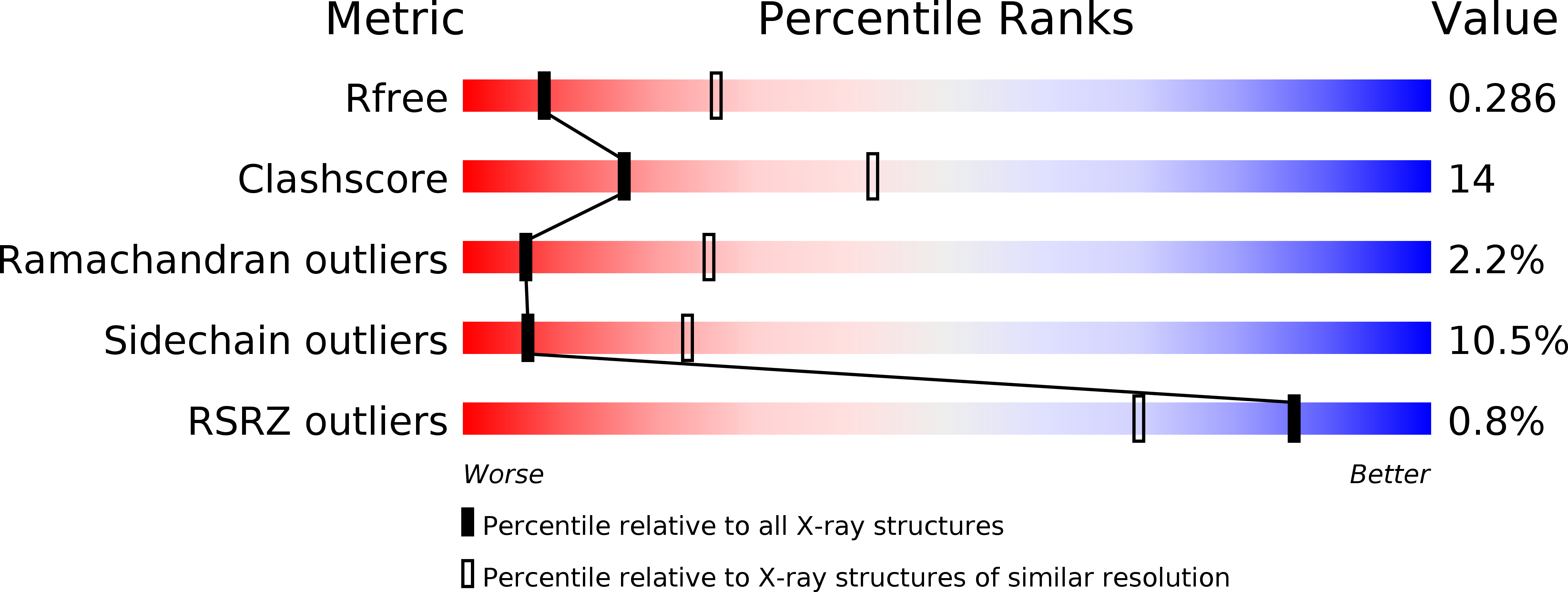
Resolution:
3.05 Å
R-Value Free:
0.28
R-Value Work:
0.21
R-Value Observed:
0.21
Space Group:
C 1 2 1
Macromolecular Entities
Polymer Type:polypeptide(L)
Molecule:HLA-DQA1 protein
Gene (Uniprot):HLA-DQA1
Chain IDs:A, C
Chain Length:213
Number of Molecules:2
Biological Source:Homo sapiens
Polymer Type:polypeptide(L)
Molecule:Peptide from Gamma-gliadin,HLA class II histocompatibility antigen, DQ beta 1 chain
Gene (Uniprot):HLA-DQB1
Mutations:Q26E, Q23E
Chain IDs:B, D
Chain Length:250
Number of Molecules:2
Biological Source:Triticum aestivum, Homo sapiens
Ligand Molecules
Primary Citation
Structural and functional studies of trans-encoded HLA-DQ2.3 (DQA1*03:01/DQB1*02:01) protein molecule
J.Biol.Chem. 287 13611 13619 (2012)
PMID: 22362761 DOI: 10.1074/jbc.M111.320374

Abstact

MHC class II molecules are composed of one α-chain and one β-chain whose membrane distal interface forms the peptide binding groove. Most of the existing knowledge on MHC class II molecules comes from the cis-encoded variants where the α- and β-chain are encoded on the same chromosome. However, trans-encoded class II MHC molecules, where the α- and β-chain are encoded on opposite chromosomes, can also be expressed. We have studied the trans-encoded class II HLA molecule DQ2.3 (DQA1*03:01/DQB1*02:01) that has received particular attention as it may explain the increased risk of certain individuals to type 1 diabetes. We report the x-ray crystal structure of this HLA molecule complexed with a gluten epitope at 3.05 Å resolution. The gluten epitope, which is the only known HLA-DQ2.3-restricted epitope, is preferentially recognized in the context of the DQ2.3 molecule by T-cell clones of a DQ8/DQ2.5 heterozygous celiac disease patient. This preferential recognition can be explained by improved HLA binding as the epitope combines the peptide-binding motif of DQ2.5 (negative charge at P4) and DQ8 (negative charge at P1). The analysis of the structure of DQ2.3 together with all other available DQ crystal structures and sequences led us to categorize DQA1 and DQB1 genes into two groups where any α-chain and β-chain belonging to the same group are expected to form a stable heterodimer.

Legend

Protein

Chemical

Disease

Primary Citation of related structures