Deposition Date
2014-11-17
Release Date
2015-03-18
Last Version Date
2023-12-20
Entry Detail
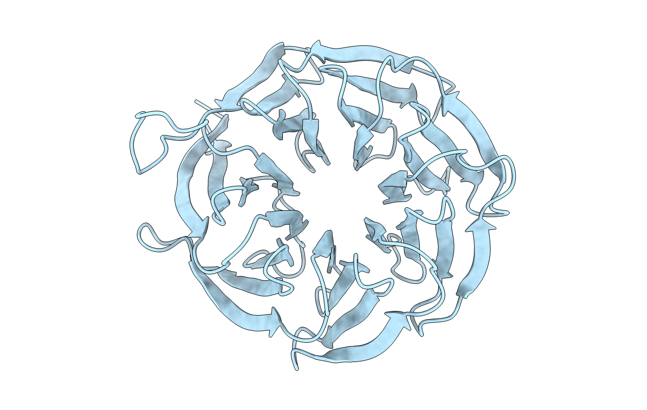
Biological Source:
Source Organism(s):
CRYPTOCOCCUS NEOFORMANS VAR. GRUBII H99 (Taxon ID: 235443)
Expression System(s):
Method Details:
Experimental Method:
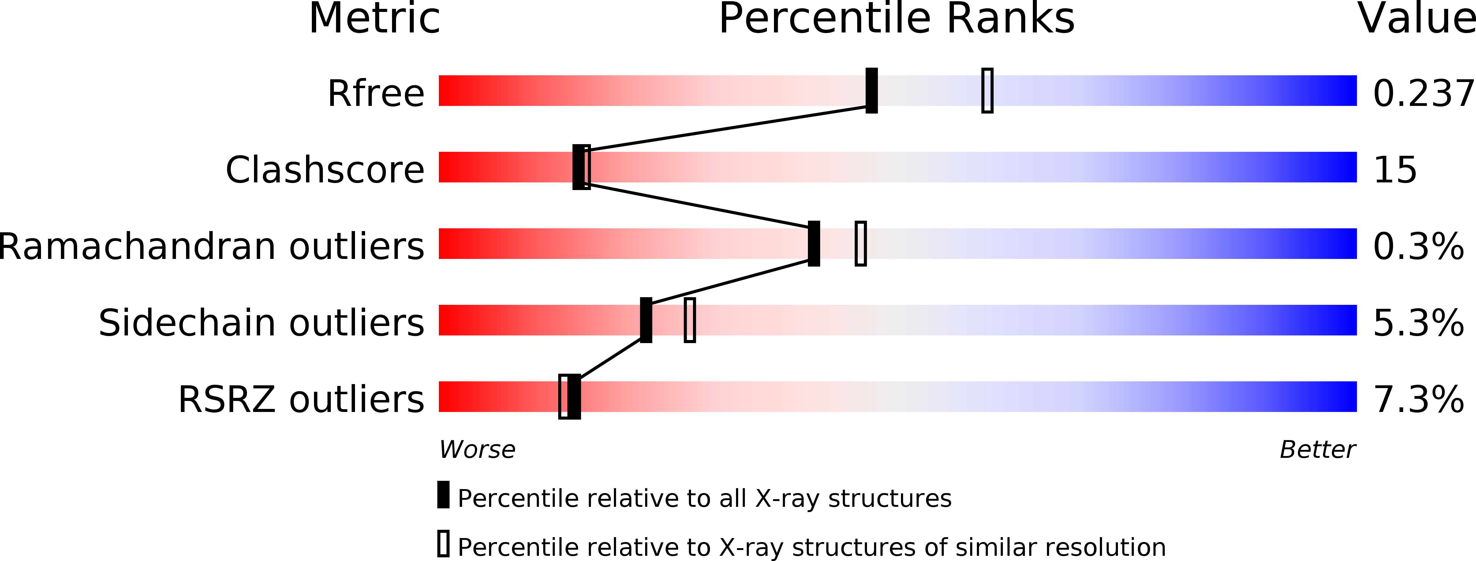
Resolution:
2.20 Å
R-Value Free:
0.23
R-Value Work:
0.19
R-Value Observed:
0.19
Space Group:
P 41 21 2