4D62 image
Deposition Date 2014-11-08
Release Date 2015-10-14
Last Version Date 2024-05-08
Entry Detail
PDB ID:
4D62
Keywords:
Title:
Structure of the carboxy-terminal domain of the turkey type 3 siadenovirus fibre, avirulent form complexed with 3-sialyllactose.
Biological Source:
Host Organism:
Method Details:
Experimental Method:
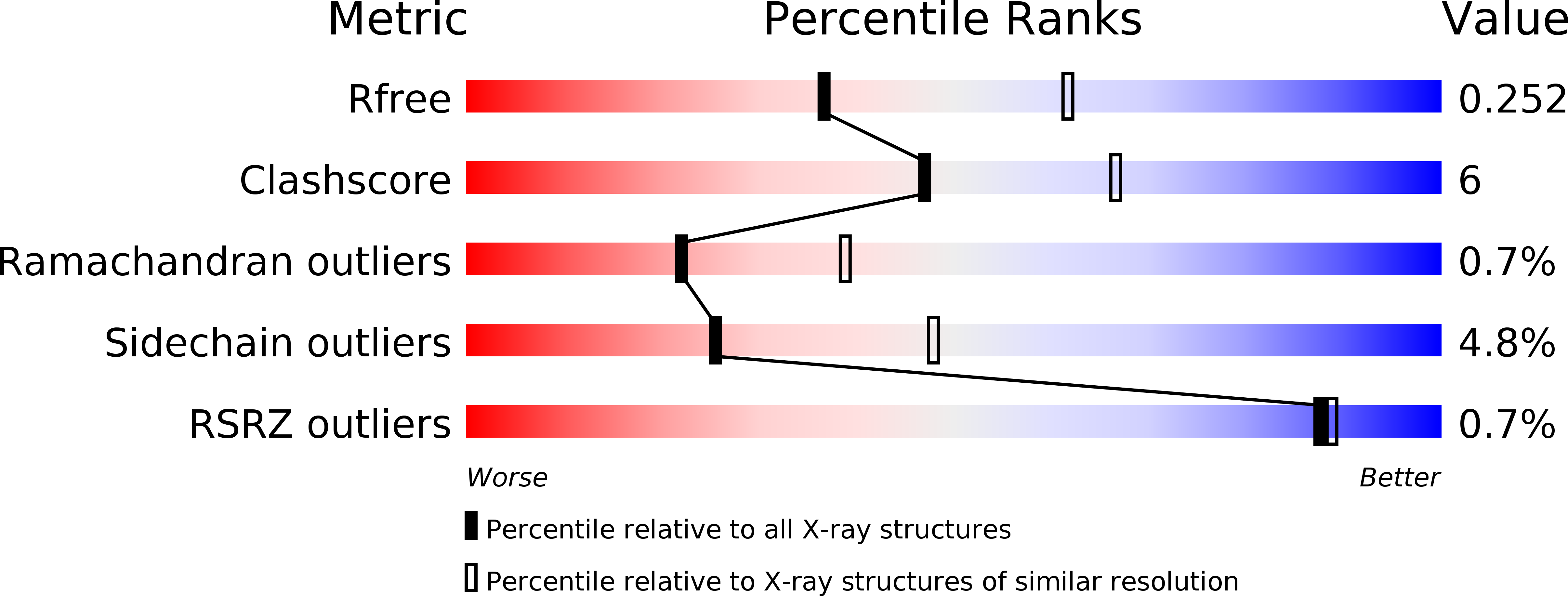
Resolution:
2.50 Å
R-Value Free:
0.25
R-Value Work:
0.19
R-Value Observed:
0.19
Space Group:
I 2 3
Macromolecular Entities
Polymer Type:polypeptide(L)
Molecule:FIBER KNOB DOMAIN
Chain IDs:A
Chain Length:187
Number of Molecules:1
Biological Source:AVIRULENT TURKEY HEMORRHAGIC ENTERITIS VIRUS
Ligand Molecules
Primary Citation
Structure and Sialyllactose Binding of the Carboxy-Terminal Head Domain of the Fibre from a Siadenovirus, Turkey Adenovirus 3.
PLoS ONE 10 e0139339 e0139339 (2015)
PMID: 26418008 DOI: 10.1371/journal.pone.0139339

Abstact

The virulent form of turkey adenovirus 3 (TAdV-3), also known as turkey hemorrhagic enteritis virus (THEV), is an economically important poultry pathogen, while the avirulent form is used as a vaccine. TAdV-3 belongs to the genus Siadenovirus. The carboxy-terminal region of its fibre does not have significant sequence similarity to any other adenovirus fibre heads of known structure. Two amino acid sequence differences between virulent and avirulent TAdV-3 map on the fibre head: where virulent TAdV-3 contains Ile354 and Thr376, avirulent TAdV-3 contains Met354 and Met376. We determined the crystal structures of the trimeric virulent and avirulent TAdV-3 fibre head domains at 2.2 Å resolution. Each monomer contains a beta-sandwich, which, surprisingly, resembles reovirus fibre head more than other adenovirus fibres, although the ABCJ-GHID topology is conserved in all. A beta-hairpin insertion in the C-strand of each trimer subunit embraces its neighbouring monomer. The avirulent and virulent TAdV-3 fibre heads are identical apart from the exact orientation of the beta-hairpin insertion. In vitro, sialyllactose was identified as a ligand by glycan microarray analysis, nuclear magnetic resonance spectroscopy, and crystallography. Its dissociation constant was measured to be in the mM range by isothermal titration calorimetry. The ligand binds to the side of the fibre head, involving amino acids Glu392, Thr419, Val420, Lys421, Asn422, and Gly423 binding to the sialic acid group. It binds slightly more strongly to the avirulent form. We propose that, in vivo, the TAdV-3 fibre may bind a sialic acid-containing cell surface component.

Legend

Protein

Chemical

Disease

Primary Citation of related structures