4D60 image
Deposition Date 2014-11-07
Release Date 2015-06-03
Last Version Date 2023-12-20
Entry Detail
PDB ID:
4D60
Title:
Structure of a dimeric Plasmodium falciparum profilin mutant
Biological Source:
Source Organism:
Method Details:
Experimental Method:
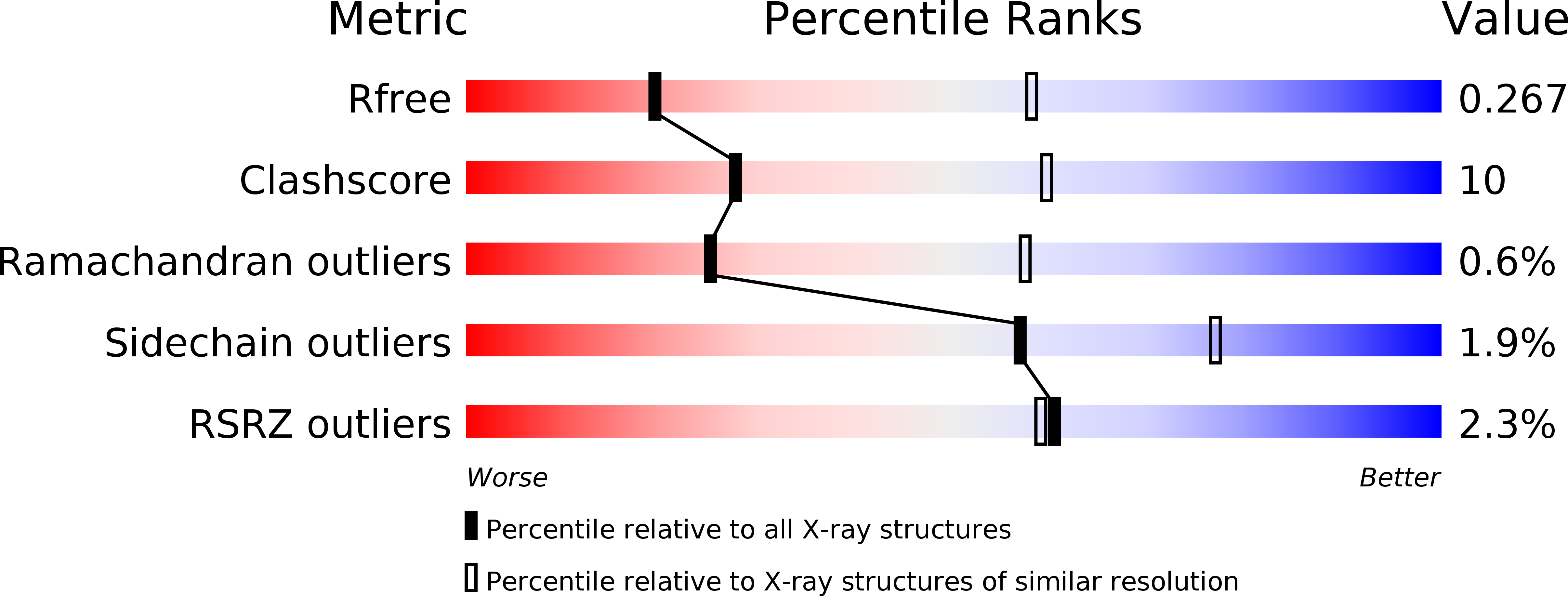
Resolution:
3.30 Å
R-Value Free:
0.26
R-Value Work:
0.23
R-Value Observed:
0.24
Space Group:
P 21 21 21
Macromolecular Entities
Polymer Type:polypeptide(L)
Molecule:PROFILIN
Chain IDs:A, B, C, D, E, F, G, H, I, J, K, L, M, N, O, P
Chain Length:169
Number of Molecules:16
Biological Source:Plasmodium falciparum
Ligand Molecules
Primary Citation
Two Independently Folding Units of Plasmodium Profilin Suggest Evolution Via Gene Fusion.
Cell.Mol.Life Sci. 72 4193 ? (2015)
PMID: 26012696 DOI: 10.1007/S00018-015-1932-0

Abstact

Gene fusion is a common mechanism of protein evolution that has mainly been discussed in the context of multidomain or symmetric proteins. Less is known about fusion of ancestral genes to produce small single-domain proteins. Here, we show with a domain-swapped mutant Plasmodium profilin that this small, globular, apparently single-domain protein consists of two foldons. The separation of binding sites for different protein ligands in the two halves suggests evolution via an ancient gene fusion event, analogous to the formation of multidomain proteins. Finally, the two fragments can be assembled together after expression as two separate gene products. The possibility to engineer both domain-swapped dimers and half-profilins that can be assembled back to a full profilin provides perspectives for engineering of novel protein folds, e.g., with different scaffolding functions.

Legend

Protein

Chemical

Disease

Primary Citation of related structures