Deposition Date
2014-10-31
Release Date
2015-04-15
Last Version Date
2024-05-08
Entry Detail
PDB ID:
4D4Z
Keywords:
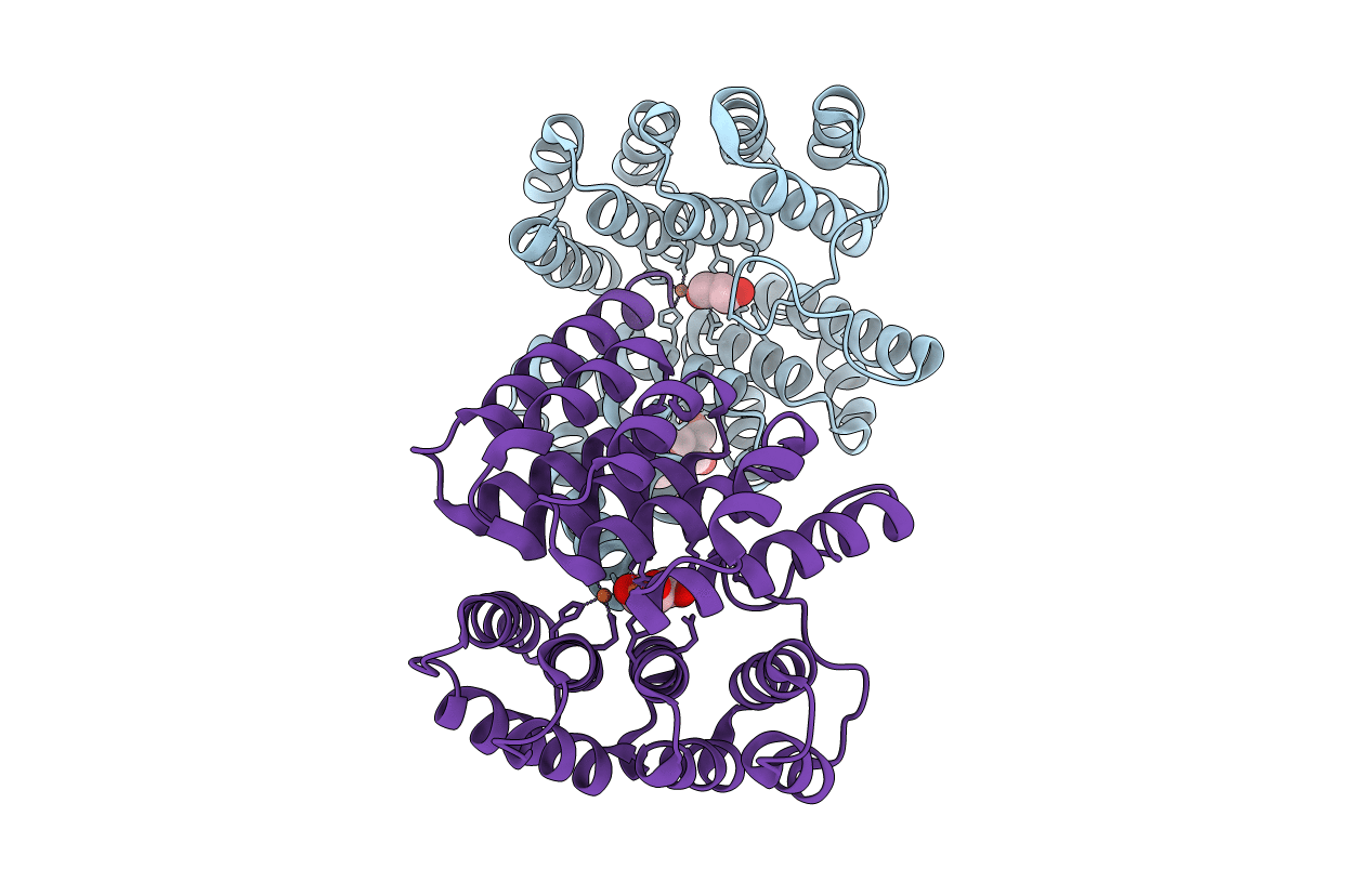
Title:
STRUCTURE OF HUMAN DEOXYHYPUSINE HYDROXYLASE in complex with glycerol
Biological Source:
Source Organism(s):
HOMO SAPIENS (Taxon ID: 9606)
Expression System(s):
Method Details:
Experimental Method:
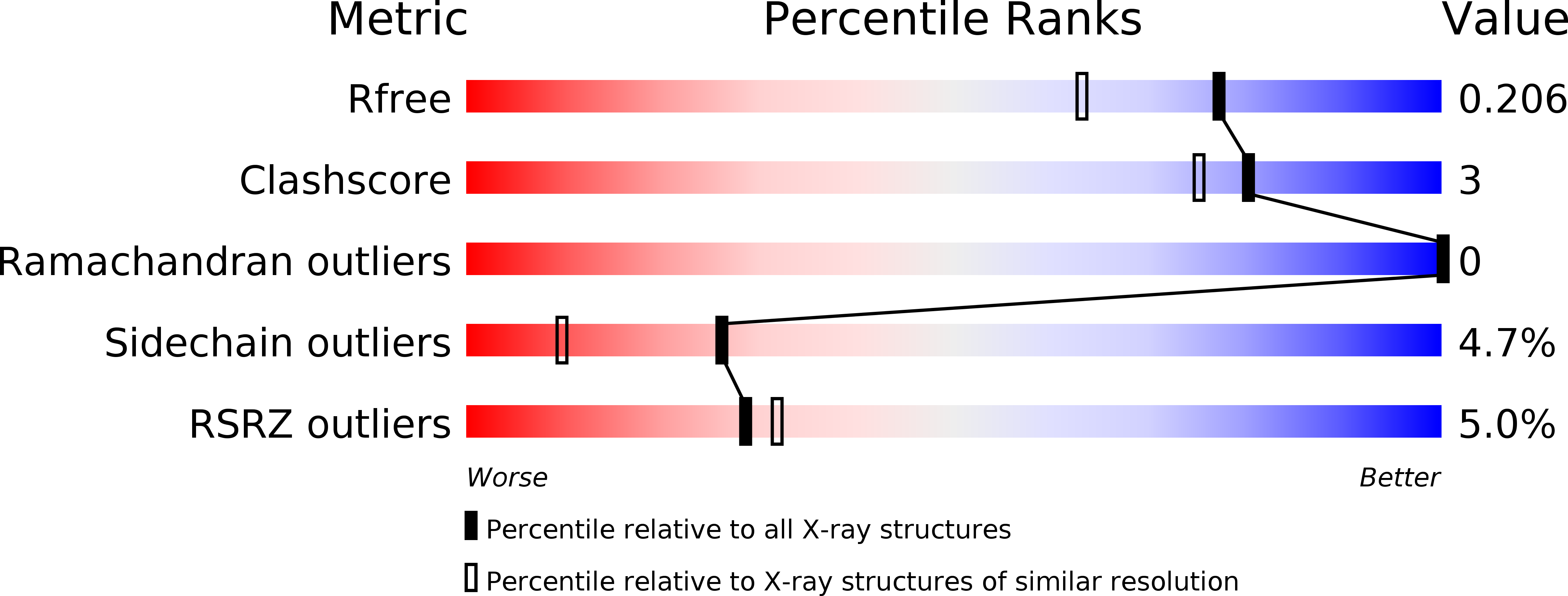
Resolution:
1.70 Å
R-Value Free:
0.19
R-Value Work:
0.17
R-Value Observed:
0.17
Space Group:
P 1 21 1