Deposition Date
2014-05-09
Release Date
2015-03-18
Last Version Date
2024-11-06
Entry Detail
PDB ID:
4D2G
Keywords:
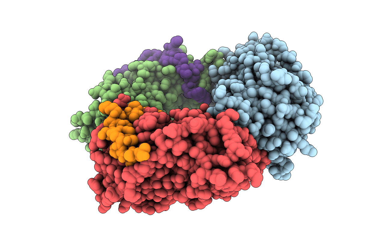
Title:
Crystal structure of human PCNA in complex with p15 peptide
Biological Source:
Source Organism(s):
HOMO SAPIENS (Taxon ID: 9606)
SYNTHETIC CONSTRUCT (Taxon ID: 32630)
SYNTHETIC CONSTRUCT (Taxon ID: 32630)
Expression System(s):
Method Details:
Experimental Method:
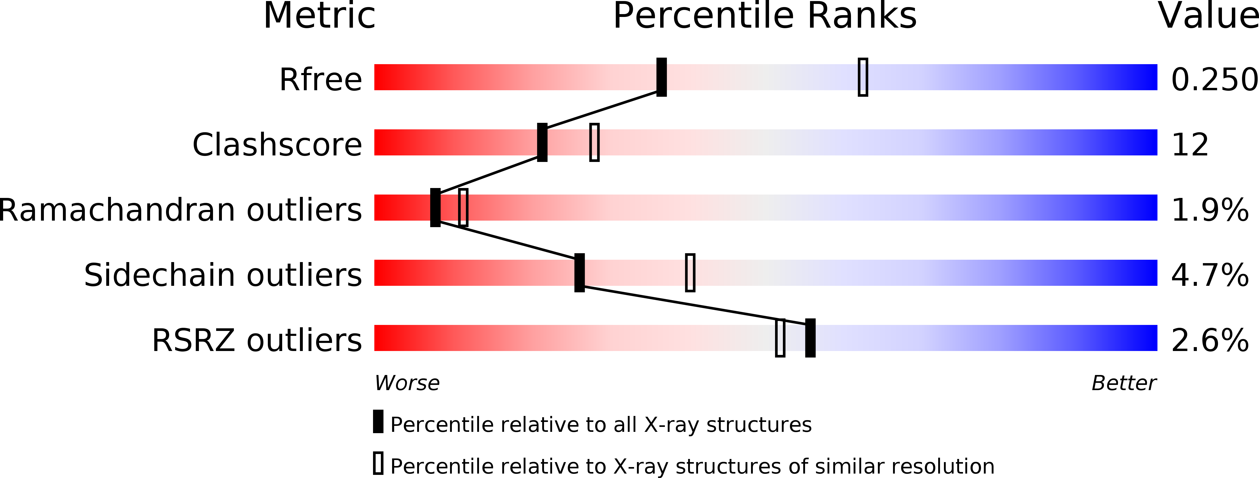
Resolution:
2.65 Å
R-Value Free:
0.24
R-Value Work:
0.17
R-Value Observed:
0.17
Space Group:
P 1 21 1