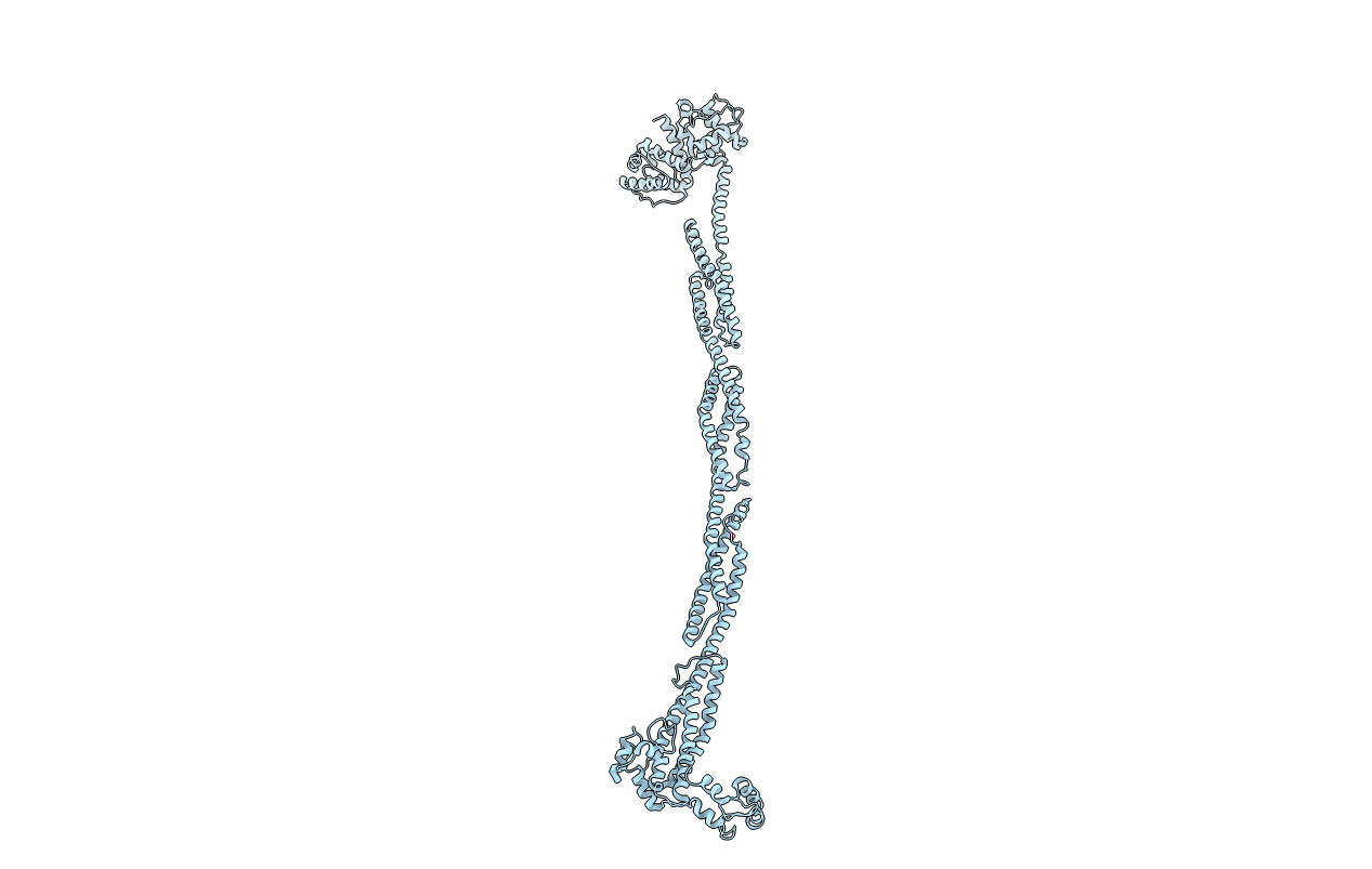
4D1E image
Entry Detail
PDB ID:
4D1E
Title:
THE CRYSTAL STRUCTURE OF HUMAN MUSCLE ALPHA-ACTININ-2
Biological Source:
Source Organism:
Host Organism:
PDB Version:
Deposition Date:
2014-05-01
Release Date:
2014-12-10
Method Details:
Experimental Method:
Resolution:
3.50 Å
R-Value Free:
0.25
R-Value Work:
0.20
R-Value Observed:
0.20
Space Group:
P 2 21 21
Macromolecular Entities
Polymer Type:polypeptide(L)
Description:ALPHA-ACTININ-2
Mutations:YES
Chain IDs:A
Chain Length:876
Number of Molecules:1
Biological Source:HOMO SAPIENS
Modified Residue
Compound ID Chain ID Parent Comp ID Details 2D Image
MLZ A LYS N-METHYL-LYSINE
Ligand Molecules
Primary Citation

Abstact

The spectrin superfamily of proteins plays key roles in assembling the actin cytoskeleton in various cell types, crosslinks actin filaments, and acts as scaffolds for the assembly of large protein complexes involved in structural integrity and mechanosensation, as well as cell signaling. α-actinins in particular are the major actin crosslinkers in muscle Z-disks, focal adhesions, and actin stress fibers. We report a complete high-resolution structure of the 200 kDa α-actinin-2 dimer from striated muscle and explore its functional implications on the biochemical and cellular level. The structure provides insight into the phosphoinositide-based mechanism controlling its interaction with sarcomeric proteins such as titin, lays a foundation for studying the impact of pathogenic mutations at molecular resolution, and is likely to be broadly relevant for the regulation of spectrin-like proteins.

Legend

Protein

Chemical

Disease

Primary Citation of related structures