Deposition Date
2014-04-30
Release Date
2014-07-23
Last Version Date
2024-05-08
Entry Detail
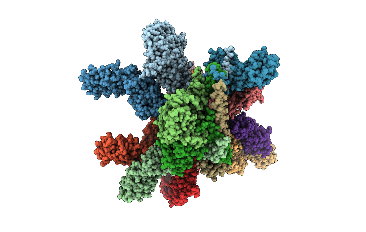
Biological Source:
Source Organism(s):
HOMO SAPIENS (Taxon ID: 9606)
Expression System(s):
Method Details:
Experimental Method:
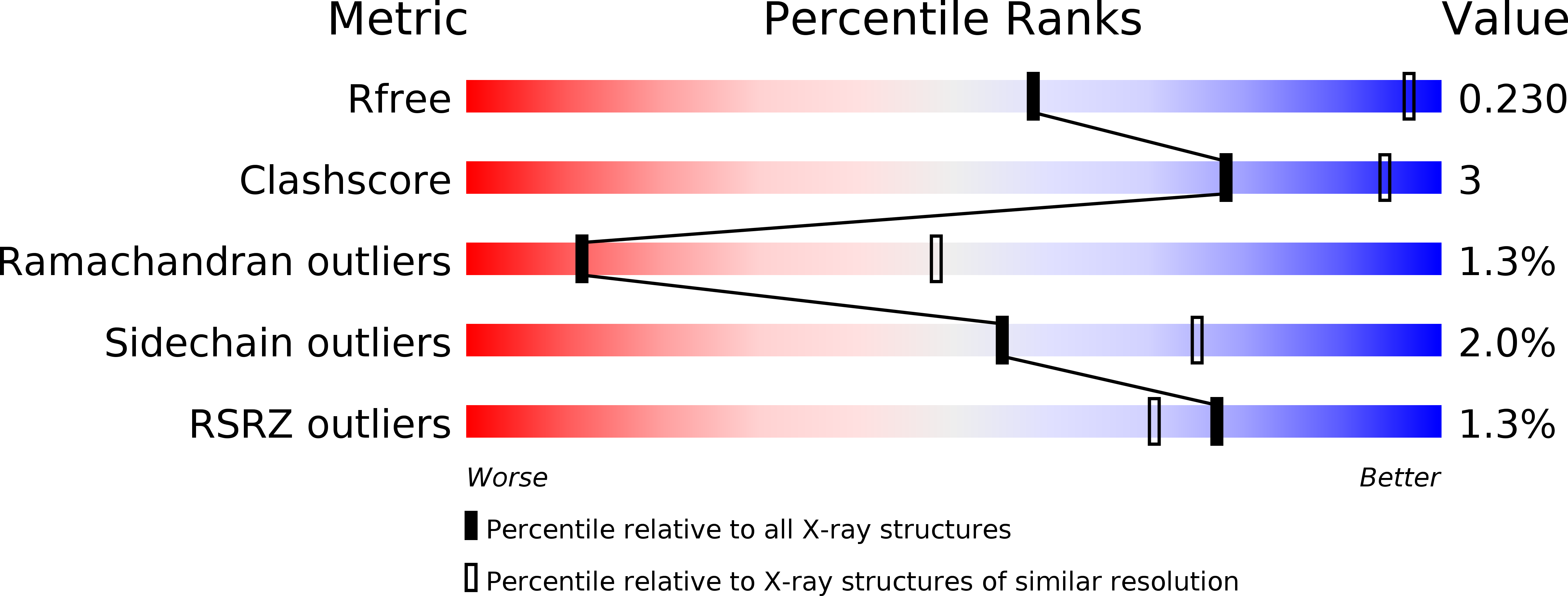
Resolution:
3.80 Å
R-Value Free:
0.22
R-Value Work:
0.19
R-Value Observed:
0.19
Space Group:
P 31