Deposition Date
2014-04-11
Release Date
2014-12-10
Last Version Date
2024-11-13
Entry Detail
PDB ID:
4CYG
Keywords:
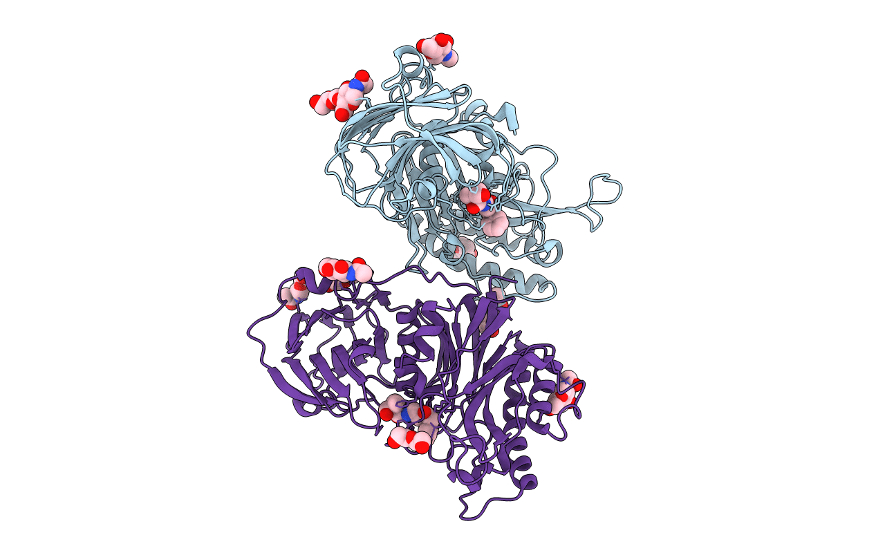
Title:
The structure of vanin-1: defining the link between metabolic disease, oxidative stress and inflammation
Biological Source:
Source Organism:
HOMO SAPIENS (Taxon ID: 9606)
Host Organism:
Method Details:
Experimental Method:
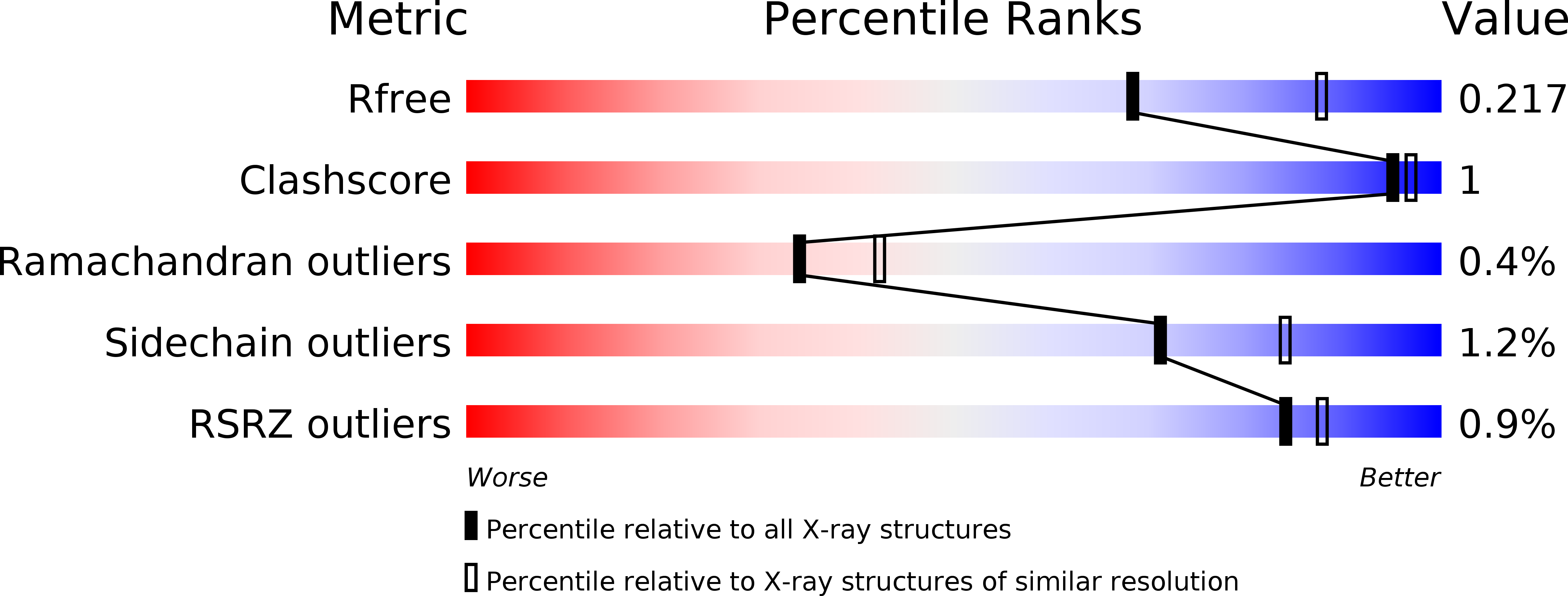
Resolution:
2.30 Å
R-Value Free:
0.21
R-Value Work:
0.18
R-Value Observed:
0.18
Space Group:
P 43 21 2