Deposition Date
2014-03-27
Release Date
2014-07-16
Last Version Date
2024-05-08
Entry Detail
PDB ID:
4CVJ
Keywords:
Title:
Neutron Structure of Compound I intermediate of Cytochrome c Peroxidase - Deuterium exchanged 100 K
Biological Source:
Source Organism(s):
SACCHAROMYCES CEREVISIAE (Taxon ID: 4932)
Expression System(s):
Method Details:
Experimental Method:
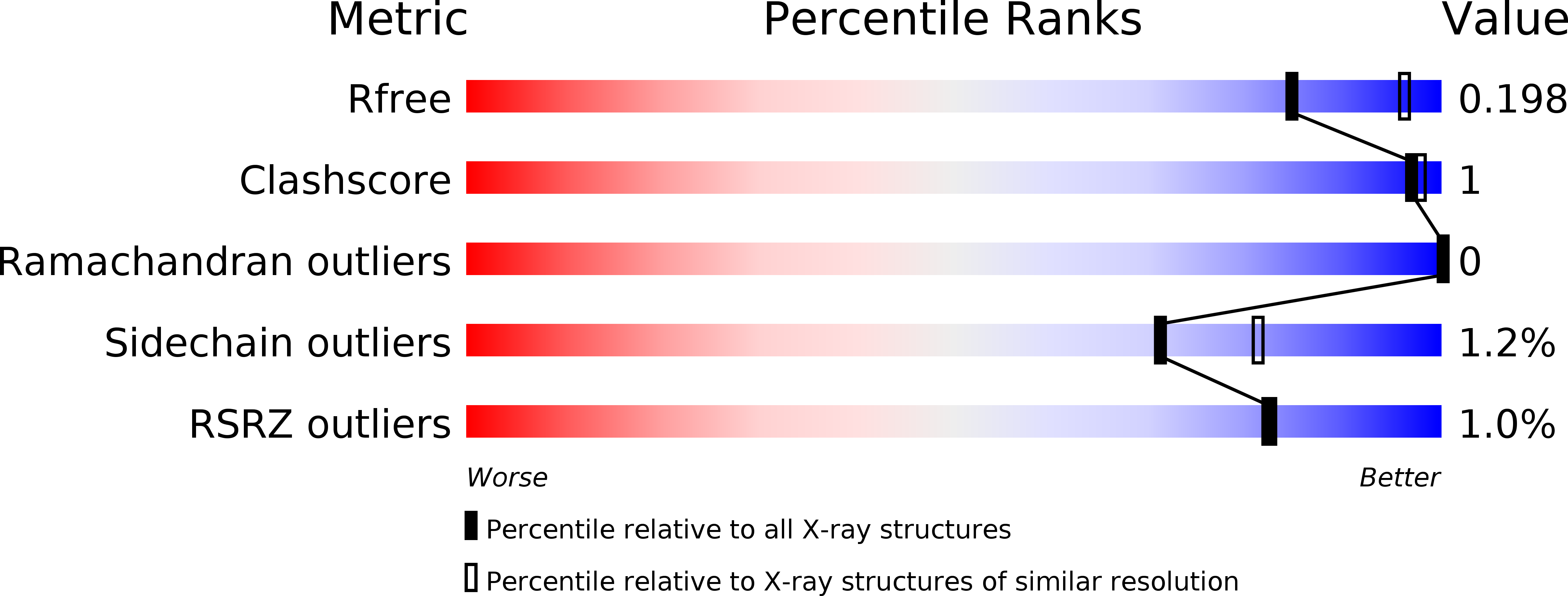
R-Value Free:
['0.20
R-Value Work:
['0.14
R-Value Observed:
['0.15
Space Group:
P 21 21 21