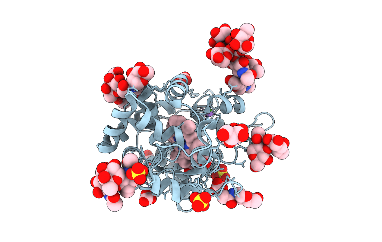
4CUO image
Entry Detail
PDB ID:
4CUO
Keywords:
Title:
Banyan peroxidase with glycosylation
Biological Source:
Source Organism:
PDB Version:
Deposition Date:
2014-03-20
Release Date:
2014-07-23
Method Details:
Experimental Method:
Resolution:
1.67 Å
R-Value Free:
0.18
R-Value Work:
0.15
R-Value Observed:
0.15
Space Group:
P 32 2 1
Macromolecular Entities
Polymer Type:polypeptide(L)
Description:BANYAN PEROXIDASE
Chain IDs:A
Chain Length:306
Number of Molecules:1
Biological Source:FICUS BENGHALENSIS
Modified Residue
Compound ID Chain ID Parent Comp ID Details 2D Image
ASN A ASN GLYCOSYLATION SITE
SNN A ASN L-3-AMINOSUCCINIMIDE
Primary Citation
Post-Translational Modification and Extended Glycosylation Pattern of a Plant Latex Peroxidase of Native Source Characterized by X-Ray Crystallography.
FEBS J. 281 4319 ? (2014)
PMID: 24980207 DOI: 10.1111/FEBS.12900

Abstact

UNLABELLED The crystal structure of banyan peroxidase purified from the latex of Ficus benghalensis has been solved at 1.67 Å resolution by single-wavelength anomalous diffraction phasing. The refined structure includes 306 amino acid residues, a heme and two calcium ions. The protein belongs to class III peroxidases and is the first one from plant latex. Extensive glycosylation was observed with N-linked glycans attached to seven asparagine residues. The enzyme is stable with respect to a wide pH range, temperature, chemical denaturants and organic solvents, probably as a result of its high glycosylation. An unexpected post-translational modification of Asp290 was identified as succinimide moiety. Kinetic parameters of banyan peroxidase have been determined using various hydrogen donor substrates and hydrogen peroxide. DATABASE Coordinates and structure factors have been deposited in the Protein Data Bank under accession number 4CUO.

Legend

Protein

Chemical

Disease

Primary Citation of related structures