4CUL image
Deposition Date 2014-03-20
Release Date 2014-05-28
Last Version Date 2024-11-06
Entry Detail
PDB ID:
4CUL
Keywords:
Title:
Structure of bovine endothelial nitric oxide synthase heme domain in complex with 6-acetyl-2-amino-7,7-dimethyl-7,8-dihydropteridin-4(3H)-one
Biological Source:
Source Organism:
BOS TAURUS (Taxon ID: 9913)
Host Organism:
Method Details:
Experimental Method:
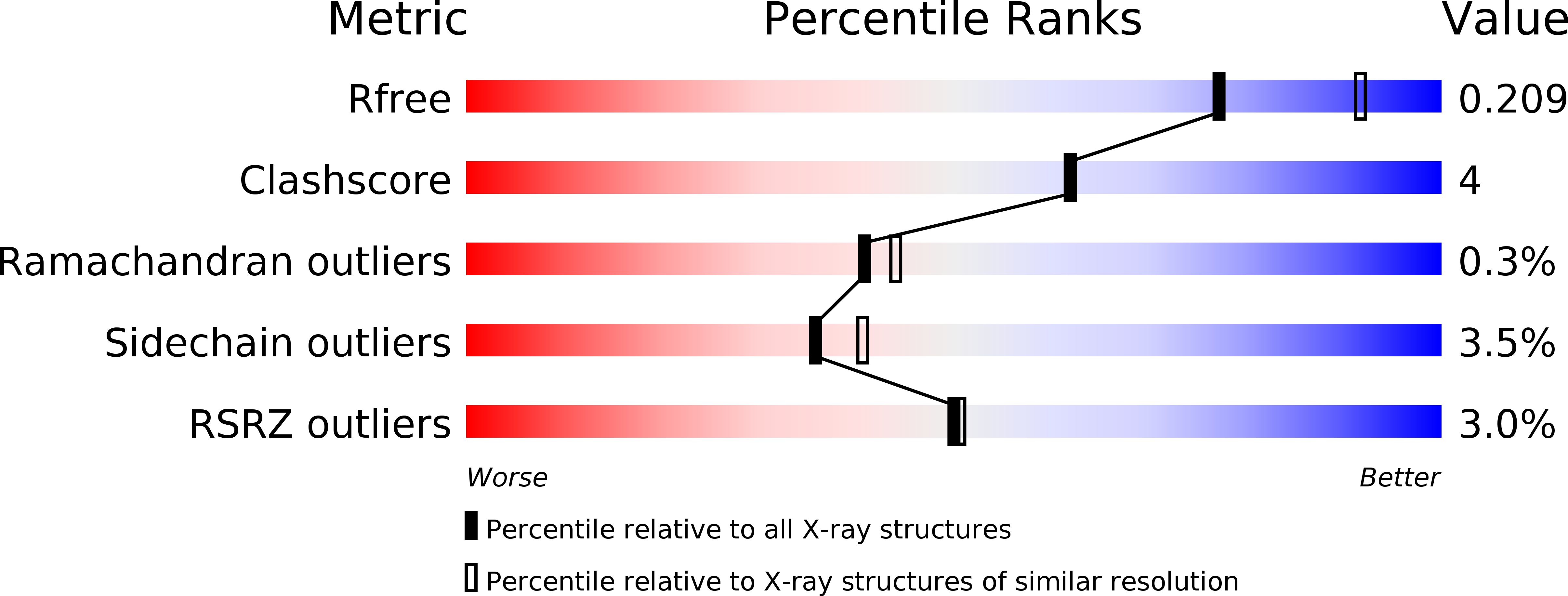
Resolution:
2.23 Å
R-Value Free:
0.20
R-Value Work:
0.16
R-Value Observed:
0.16
Space Group:
P 21 21 21
Macromolecular Entities
Polymer Type:polypeptide(L)
Molecule:NITRIC OXIDE SYNTHASE, ENDOTHELIAL
Gene (Uniprot):NOS3
Chain IDs:A, B
Chain Length:443
Number of Molecules:2
Biological Source:BOS TAURUS
Modified Residue
Compound ID Chain ID Parent Comp ID Details 2D Image
CAS A CYS S-(DIMETHYLARSENIC)CYSTEINE
Primary Citation
Communication between the Zinc and Tetrahydrobiopterin Binding Sites in Nitric Oxide Synthase.
Biochemistry 53 4216 ? (2014)
PMID: 24819538 DOI: 10.1021/BI5003986

Abstact

The nitric oxide synthase (NOS) dimer is stabilized by a Zn(2+) ion coordinated to four symmetry-related Cys residues exactly along the dimer 2-fold axis. Each of the two essential tetrahydrobiopterin (H4B) molecules in the dimer interacts directly with the heme, and each H4B molecule is ~15 Å from the Zn(2+). We have determined the crystal structures of the bovine endothelial NOS dimer oxygenase domain bound to three different pterin analogues, which reveal an intimate structural communication between the H4B and Zn(2+) sites. The binding of one of these compounds, 6-acetyl-2-amino-7,7-dimethyl-7,8-dihydro-4(3H)-pteridinone (1), to the pterin site and Zn(2+) binding are mutually exclusive. Compound 1 both directly and indirectly disrupts hydrogen bonding between key residues in the Zn(2+) binding motif, resulting in destabilization of the dimer and a complete disruption of the Zn(2+) site. Addition of excess Zn(2+) stabilizes the Zn(2+) site at the expense of weakened binding of 1. The unique structural features of 1 that disrupt the dimer interface are extra methyl groups that extend into the dimer interface and force a slight opening of the dimer, thus resulting in disruption of the Zn(2+) site. These results illustrate a very delicate balance of forces and structure at the dimer interface that must be maintained to properly form the Zn(2+), pterin, and substrate binding sites.

Legend

Protein

Chemical

Disease

Primary Citation of related structures