4CSD image
Deposition Date 2014-03-06
Release Date 2014-10-08
Last Version Date 2023-12-20
Entry Detail
PDB ID:
4CSD
Title:
Structure of Monomeric Ralstonia solanacearum lectin
Biological Source:
Source Organism:
Host Organism:
Method Details:
Experimental Method:
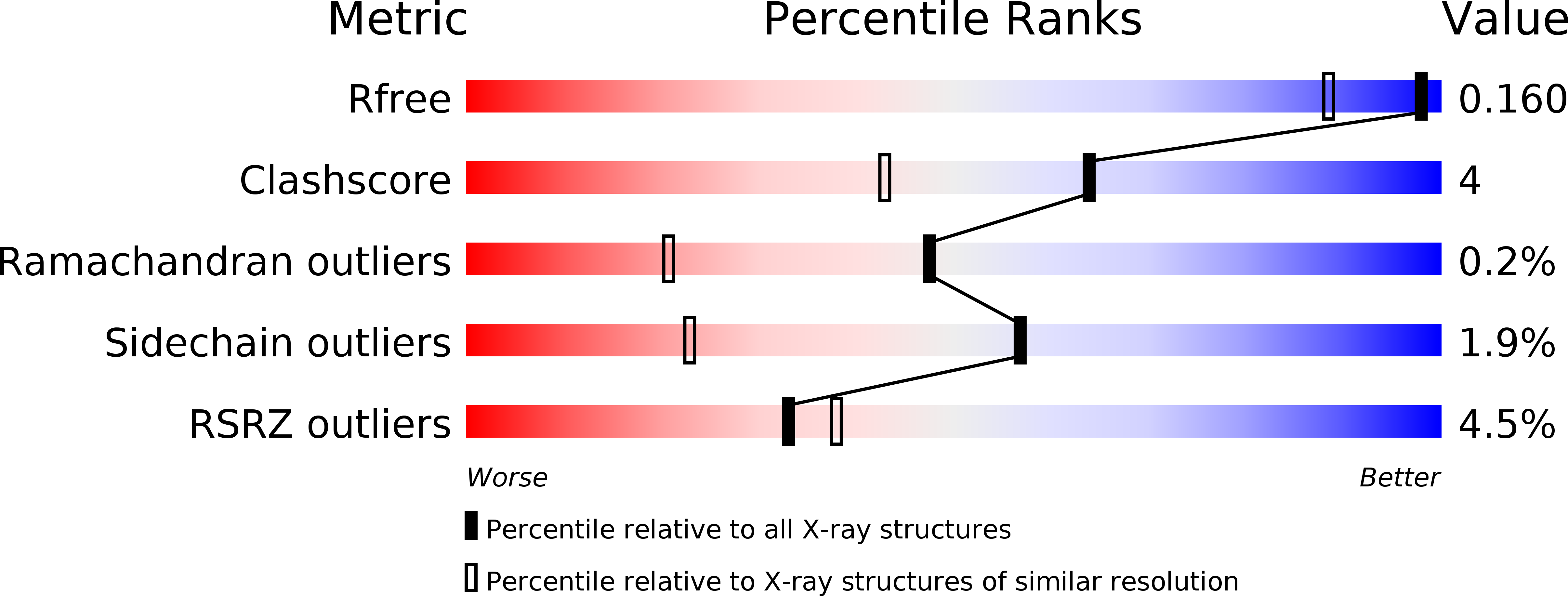
Resolution:
1.35 Å
R-Value Free:
0.14
R-Value Work:
0.11
R-Value Observed:
0.11
Space Group:
P 1 21 1
Macromolecular Entities
Structures with similar UniProt ID
Protein Blast
Polymer Type:polypeptide(L)
Molecule:FUCOSE-BINDING LECTIN PROTEIN
Gene (Uniprot):rsl
Chain IDs:A, B
Chain Length:272
Number of Molecules:2
Biological Source:RALSTONIA SOLANACEARUM
Primary Citation
Membrane Deformation by Neolectins with Engineered Glycolipid Binding Sites.
Angew.Chem.Int.Ed.Engl. 53 9267 ? (2014)
PMID: 25044646 DOI: 10.1002/ANIE.201404568

Abstact

Lectins are glycan-binding proteins that are involved in the recognition of glycoconjugates at the cell surface. When binding to glycolipids, multivalent lectins can affect their distribution and alter membrane shapes. Neolectins have now been designed with controlled number and position of binding sites to decipher the role of multivalency on avidity to a glycosylated surface and on membrane dynamics of glycolipids. A monomeric hexavalent neolectin has been first engineered from a trimeric hexavalent bacterial lectin, From this neolectin template, 13 different neolectins with a valency ranging from 0 to 6 were designed, produced, and analyzed for their ability to bind fucose in solution, to attach to a glycosylated surface and to invaginate glycolipid-containing giant liposomes. Whereas the avidity only depends on the presence of at least two binding sites, the ability to bend and invaginate membranes critically depends on the distance between two adjacent binding sites.

Legend

Protein

Chemical

Disease

Primary Citation of related structures