Deposition Date
1989-10-18
Release Date
1990-10-15
Last Version Date
2024-10-23
Entry Detail
PDB ID:
4CPV
Keywords:
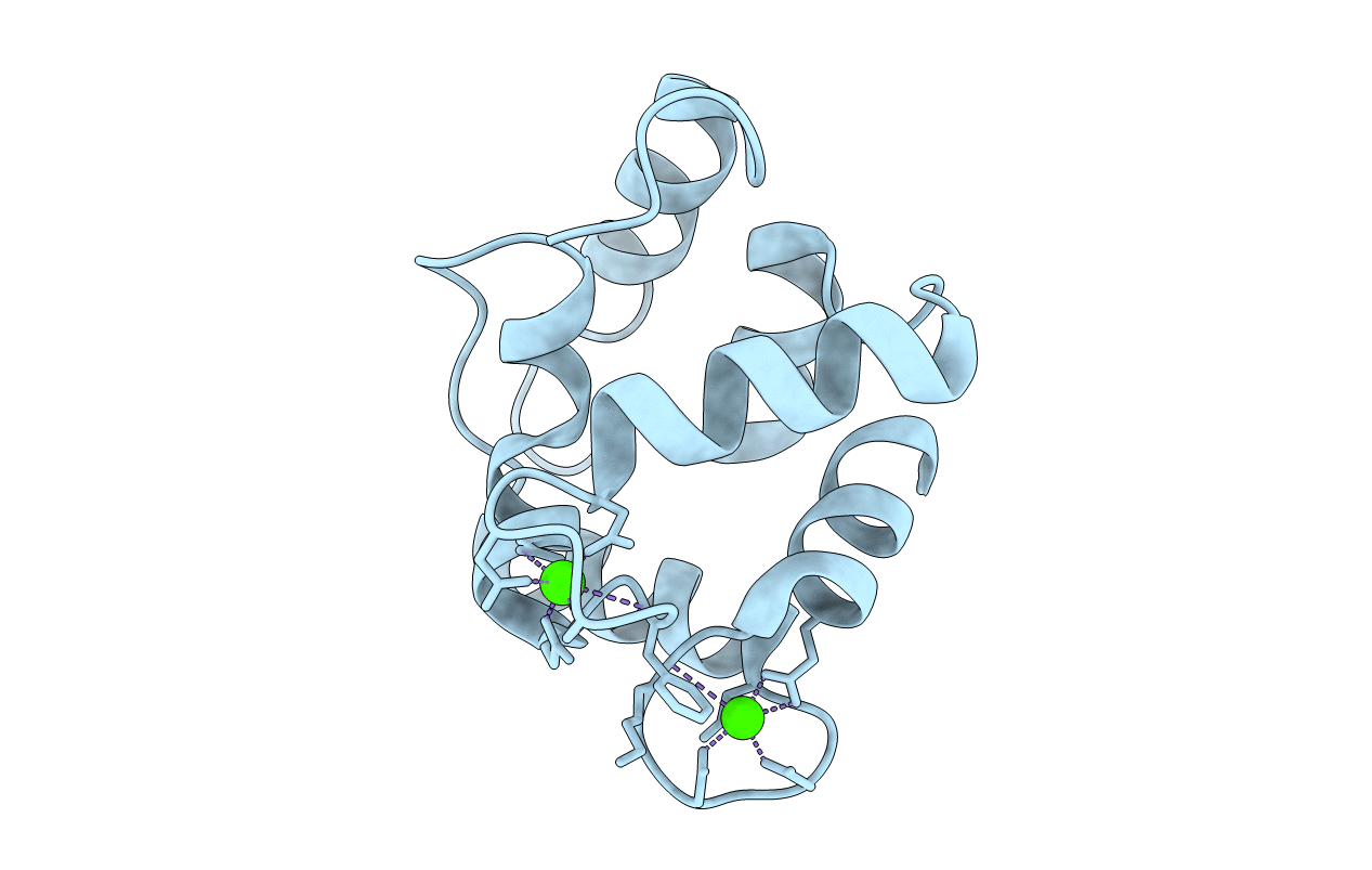
Title:
REFINED CRYSTAL STRUCTURE OF CALCIUM-LIGANDED CARP PARVALBUMIN 4.25 AT 1.5-ANGSTROMS RESOLUTION
Biological Source:
Source Organism(s):
Cyprinus carpio (Taxon ID: 7962)
Method Details:
Experimental Method:
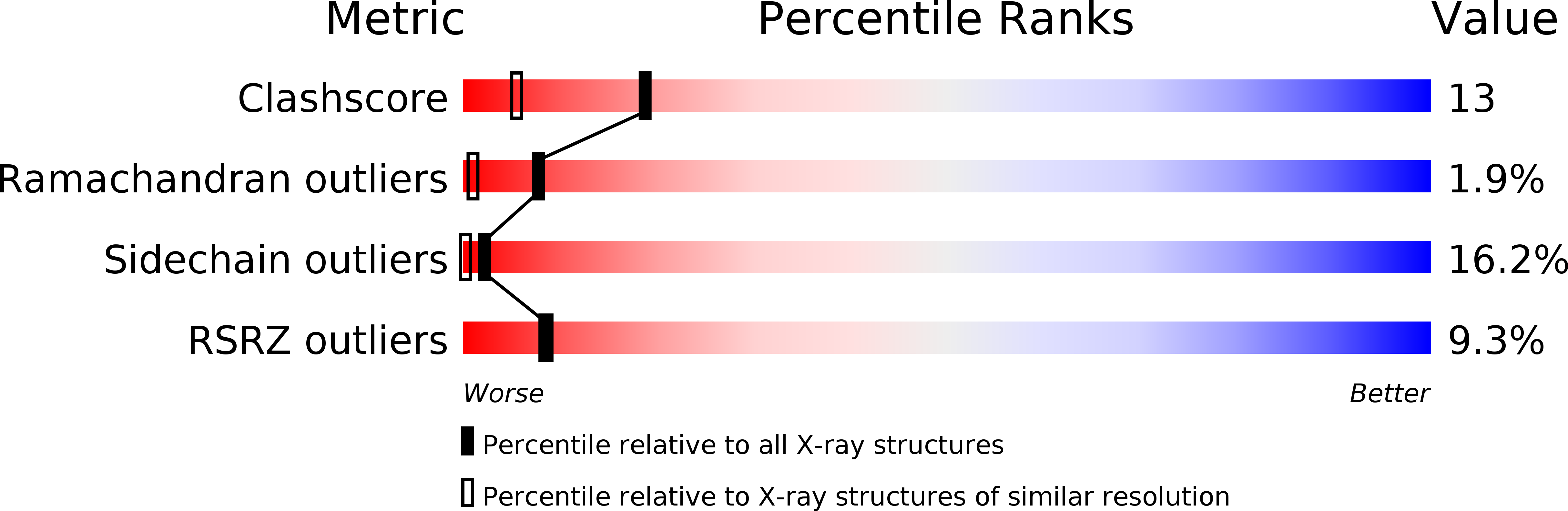
Resolution:
1.50 Å
R-Value Observed:
0.21
Space Group:
C 1 2 1