
Deposition Date
2014-01-21
Release Date
2015-02-18
Last Version Date
2023-12-20
Entry Detail
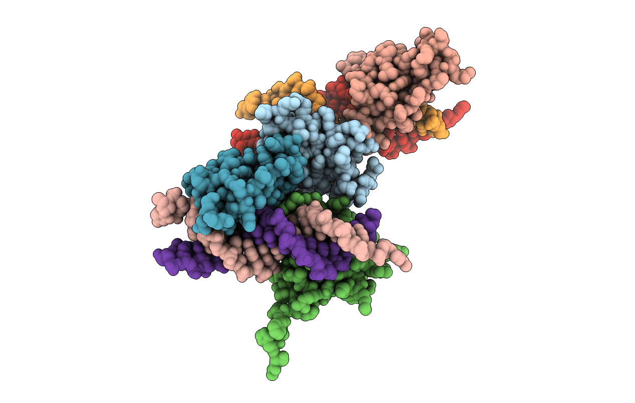
PDB ID:
4CN3
Keywords:
Title:
Crystal Structure of the Human Retinoid X Receptor DNA-Binding Domain Bound to the Human Gde1SpA Response Element
Biological Source:
Source Organism:
HOMO SAPIENS (Taxon ID: 9606)
Host Organism:
Method Details:
Experimental Method:
Resolution:
2.35 Å
R-Value Free:
0.24
R-Value Work:
0.17
R-Value Observed:
0.18
Space Group:
P 21 21 21
