Deposition Date
2013-12-19
Release Date
2014-01-22
Last Version Date
2023-12-20
Entry Detail
PDB ID:
4CJ7
Keywords:
Title:
Structure of Crenactin, an archeal actin-like protein
Biological Source:
Source Organism:
PYROBACULUM CALIDIFONTIS (Taxon ID: 410359)
Host Organism:
Method Details:
Experimental Method:
Resolution:
3.20 Å
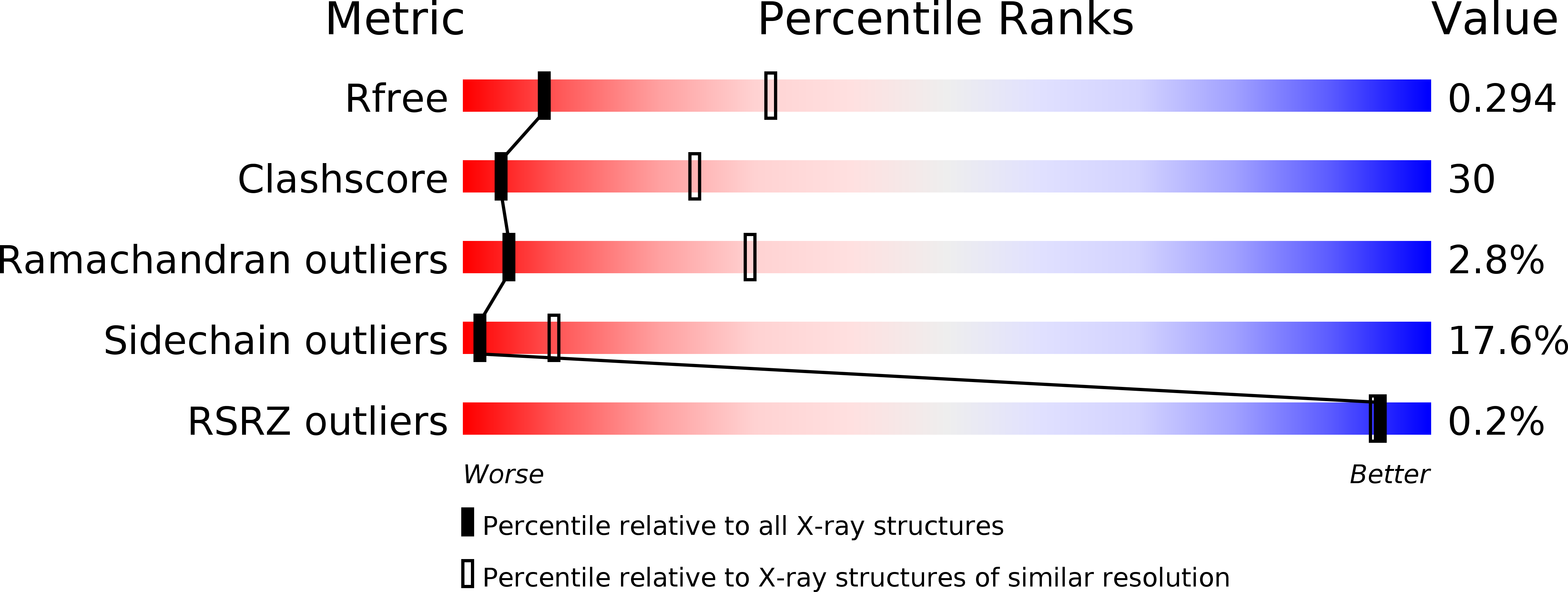
R-Value Free:
0.29
R-Value Work:
0.23
R-Value Observed:
0.23
Space Group:
P 41 21 2