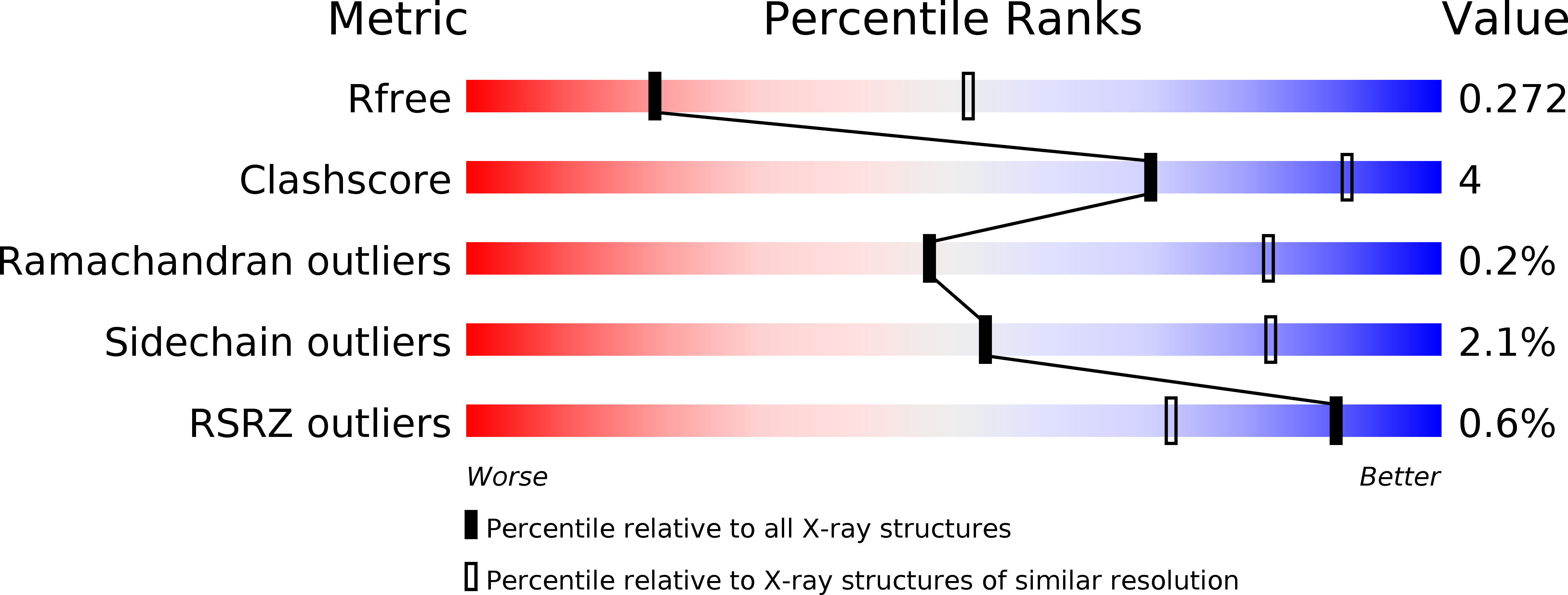
4CID image
Deposition Date 2013-12-06
Release Date 2014-02-19
Last Version Date 2024-11-13
Entry Detail
PDB ID:
4CID
Keywords:
Title:
Structural insights into the N-terminus of the EHD2 ATPase
Biological Source:
Source Organism:
MUS MUSCULUS (Taxon ID: 10090)
Host Organism:
Method Details:
Experimental Method:
Resolution:
3.00 Å
R-Value Free:
0.27
R-Value Work:
0.23
R-Value Observed:
0.23
Space Group:
C 1 2 1
Macromolecular Entities
Polymer Type:polypeptide(L)
Molecule:EH DOMAIN-CONTAINING PROTEIN 2
Gene (Uniprot):Ehd2
Mutations:YES
Chain IDs:A
Chain Length:550
Number of Molecules:1
Biological Source:MUS MUSCULUS
Modified Residue
Compound ID Chain ID Parent Comp ID Details 2D Image
MSE A MET SELENOMETHIONINE
Primary Citation
Structural Insights Into Membrane Interaction and Caveolar Targeting of Dynamin-Like Ehd2.
Structure 22 409 ? (2014)
PMID: 24508342 DOI: 10.1016/J.STR.2013.12.015

Abstact

The dynamin-related Eps15-homology domain-containing protein 2 (EHD2) is a membrane-remodeling ATPase that regulates the dynamics of caveolae. Here, we established an electron paramagnetic resonance (EPR) approach to characterize structural features of membrane-bound EHD2. We show that residues at the tip of the helical domain can insert into the membrane and may create membrane curvature by a wedging mechanism. Using EPR and X-ray crystallography, we found that the N terminus is folded into a hydrophobic pocket of the GTPase domain in solution and can be released into the membrane. Cryoelectron microscopy demonstrated that the N terminus is not essential for oligomerization of EHD2 into a membrane-anchored scaffold. Instead, we found a function of the N terminus in regulating targeting and stable association of EHD2 to caveolae. Our data uncover an unexpected, membrane-induced regulatory switch in EHD2 and demonstrate the versatility of EPR to study structure and function of dynamin superfamily proteins.

Legend

Protein

Chemical

Disease

Primary Citation of related structures