4CI6 image
Deposition Date 2013-12-06
Release Date 2015-01-28
Last Version Date 2023-12-20
Entry Detail
PDB ID:
4CI6
Title:
Mechanisms of crippling actin-dependent phagocytosis by YopO
Biological Source:
Source Organism:
Host Organism:
Method Details:
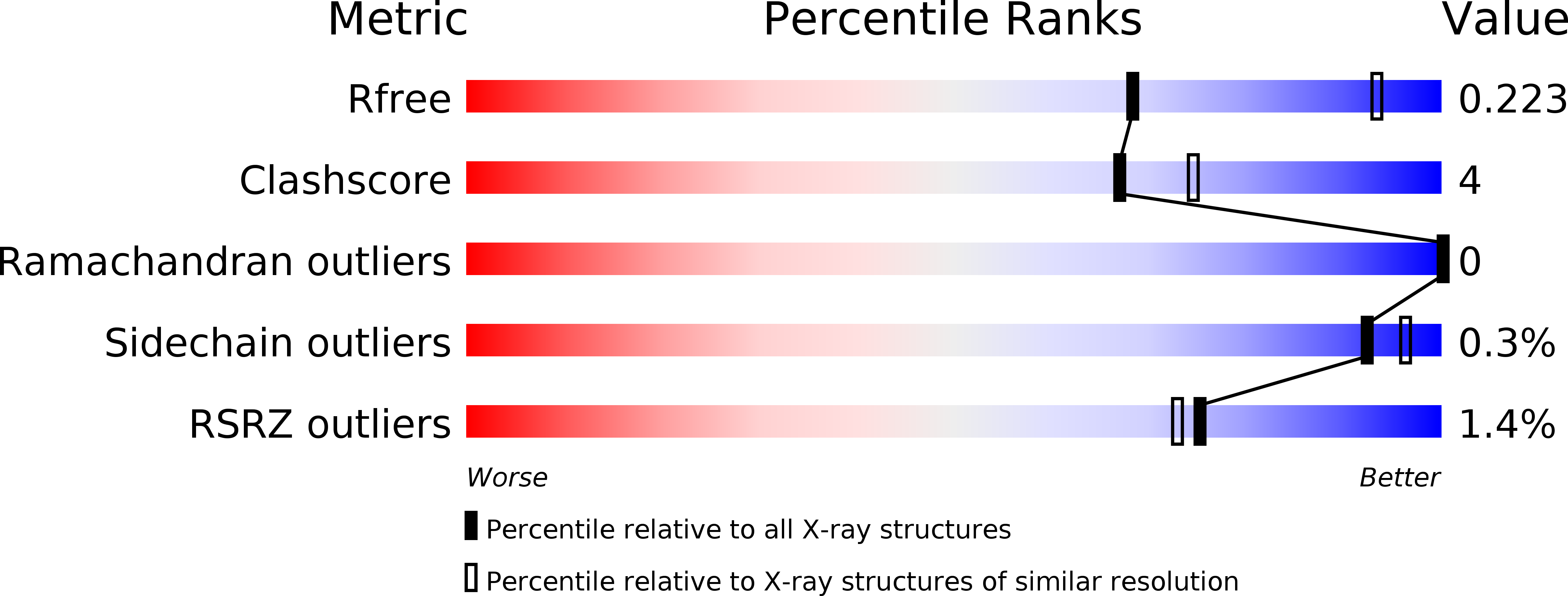
Experimental Method:
Resolution:
2.65 Å
R-Value Free:
0.22
R-Value Work:
0.18
R-Value Observed:
0.18
Space Group:
P 1 21 1
Macromolecular Entities
Polymer Type:polypeptide(L)
Molecule:ACTIN
Chain IDs:A
Chain Length:376
Number of Molecules:1
Biological Source:SPODOPTERA FRUGIPERDA
Polymer Type:polypeptide(L)
Molecule:PROTEIN KINASE YOPO
Gene (Uniprot):yopO
Mutations:YES
Chain IDs:B
Chain Length:643
Number of Molecules:1
Biological Source:YERSINIA ENTEROCOLITICA
Primary Citation
Yersinia Effector Yopo Uses Actin as Bait to Phosphorylate Proteins that Regulate Actin Polymerization.
Nat.Struct.Mol.Biol. 22 248 ? (2015)
PMID: 25664724 DOI: 10.1038/NSMB.2964

Abstact

Pathogenic Yersinia species evade host immune systems through the injection of Yersinia outer proteins (Yops) into phagocytic cells. One Yop, YopO, also known as YpkA, induces actin-filament disruption, impairing phagocytosis. Here we describe the X-ray structure of Yersinia enterocolitica YopO in complex with actin, which reveals that YopO binds to an actin monomer in a manner that blocks polymerization yet allows the bound actin to interact with host actin-regulating proteins. SILAC-MS and biochemical analyses confirm that actin-polymerization regulators such as VASP, EVL, WASP, gelsolin and the formin diaphanous 1 are directly sequestered and phosphorylated by YopO through formation of ternary complexes with actin. This leads to a model in which YopO at the membrane sequesters actin from polymerization while using the bound actin as bait to recruit, phosphorylate and misregulate host actin-regulating proteins to disrupt phagocytosis.

Legend

Protein

Chemical

Disease

Primary Citation of related structures