4CHG image
Deposition Date 2013-12-02
Release Date 2014-11-12
Last Version Date 2024-10-23
Entry Detail
PDB ID:
4CHG
Keywords:
Title:
Crystal structure of VapBC15 complex from Mycobacterium tuberculosis
Biological Source:
Source Organism:
Method Details:
Experimental Method:
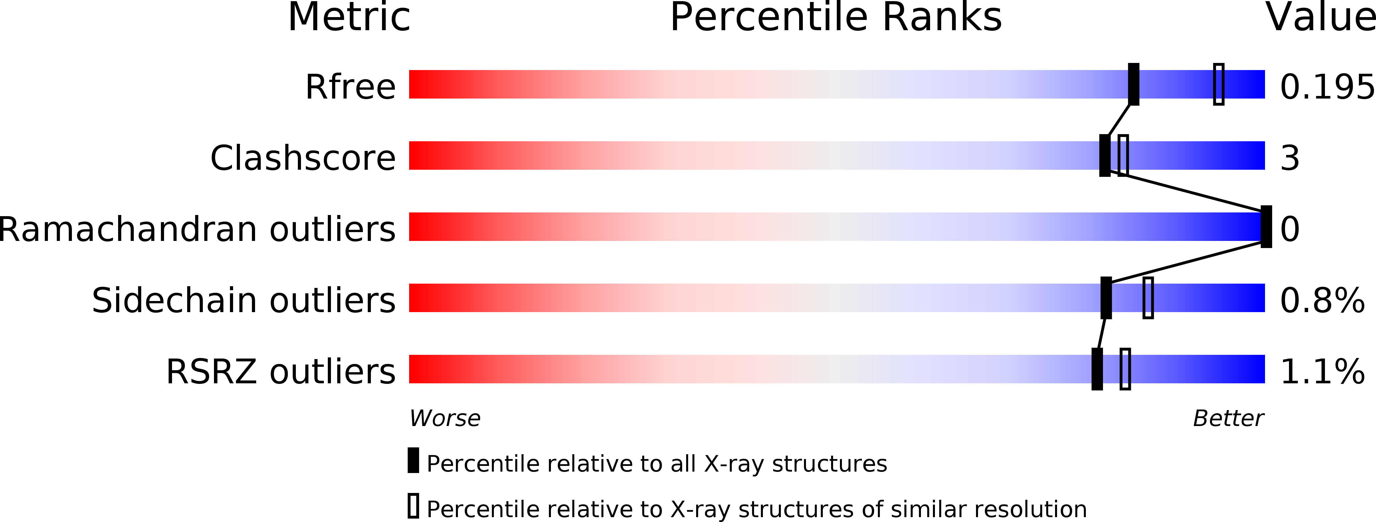
Resolution:
2.10 Å
R-Value Free:
0.19
R-Value Work:
0.16
R-Value Observed:
0.16
Space Group:
P 21 21 21
Macromolecular Entities
Polymer Type:polypeptide(L)
Molecule:PROBABLE RIBONUCLEASE VAPC15
Chain IDs:A, B, C, D, E, F
Chain Length:133
Number of Molecules:6
Biological Source:MYCOBACTERIUM TUBERCULOSIS
Polymer Type:polypeptide(L)
Molecule:ANTITOXIN VAPB15
Chain IDs:G, H, I, J
Chain Length:88
Number of Molecules:4
Biological Source:MYCOBACTERIUM TUBERCULOSIS
Modified Residue
Compound ID Chain ID Parent Comp ID Details 2D Image
MSE A MET SELENOMETHIONINE
Primary Citation
Crystal Structure of the Vapbc-15 Complex from Mycobacterium Tuberculosis Reveals a Two-Metal Ion Dependent Pin-Domain Ribonuclease And a Variable Mode of Toxin-Antitoxin Assembly.
J.Struct.Biol. 188 249 ? (2014)
PMID: 25450593 DOI: 10.1016/J.JSB.2014.10.002

Abstact

Although PIN (PilT N-terminal)-domain proteins are known to have ribonuclease activity, their specific mechanism of action remains unknown. VapCs form a family of ribonucleases that possess a PIN-domain assembly and are known as toxins. The activities of VapCs are impaired by VapB antitoxins. Here we present the crystal structure of the VapBC-15 toxin-antitoxin complex from Mycobacterium tuberculosis determined to 2.1Å resolution. The VapB-15 and VapC-15 components assemble into one heterotetramer (VapB2C2) and two heterotrimers (VapBC2) in each asymmetric unit of the crystal. The active site of VapC-15 toxin consists of a cluster of acidic amino acid residues and two divalent metal ions, forming a well organised ribonuclease active site. The distribution of the catalytic-site residues of the VapC-15 toxin is similar to that of T4 RNase H and of Methanococcus jannaschii FEN-1, providing strong evidence that these three proteins share a similar mechanism of activity. The presence of both VapB2C2 and VapBC2 emphasizes the fact that the same antitoxin can bind the toxin in 1:1 and 1:2 ratios. The crystal structure determination of the VapBC-15 complex reveals for the first time a PIN-domain ribonuclease protein that shows two metal ions at the active site and a variable mode of toxin-antitoxin assembly. The structure further shows that VapB-15 antitoxin binds to the same groove meant for the binding of putative substrate (RNA), resulting in the inhibition of VapC-15's toxicity.

Legend

Protein

Chemical

Disease

Primary Citation of related structures