4CH2 image
Deposition Date 2013-11-28
Release Date 2013-12-11
Last Version Date 2024-10-23
Entry Detail
PDB ID:
4CH2
Title:
Low-salt crystal structure of a thrombin-GpIbalpha peptide complex
Biological Source:
Source Organism:
HOMO SAPIENS (Taxon ID: 9606)
Host Organism:
Method Details:
Experimental Method:
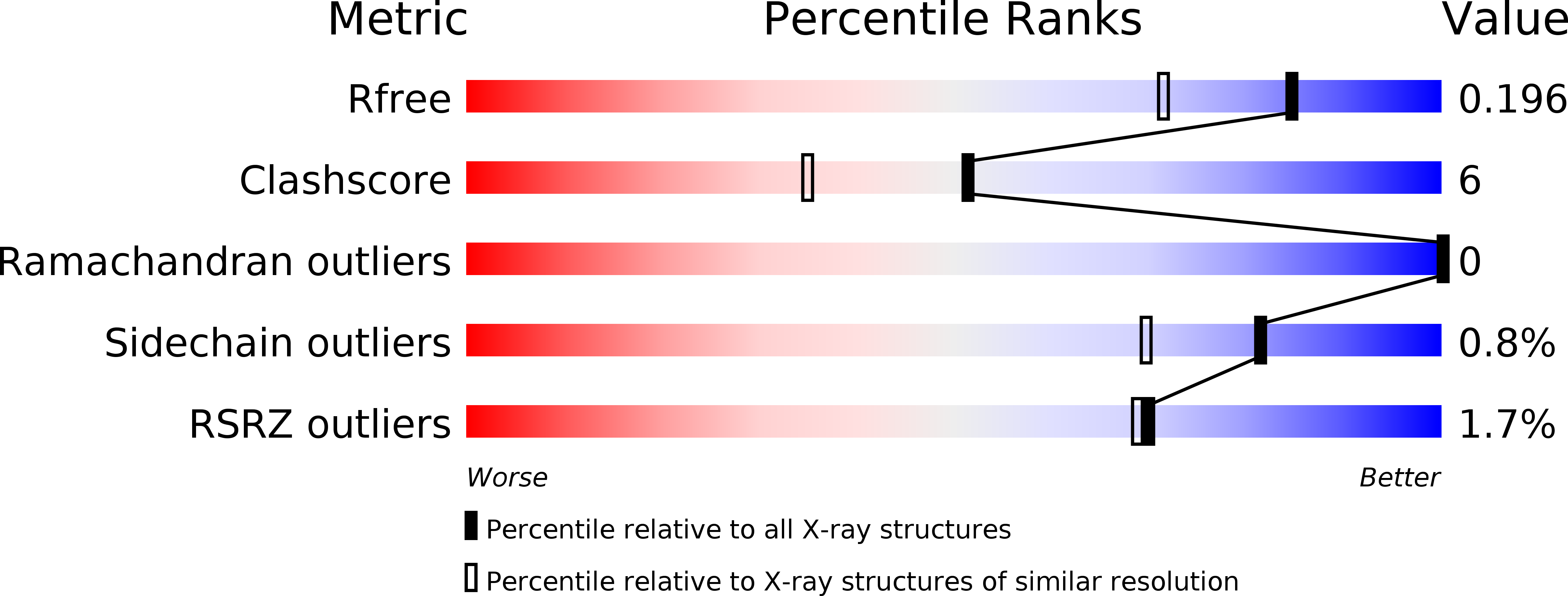
Resolution:
1.60 Å
R-Value Free:
0.18
R-Value Work:
0.15
R-Value Observed:
0.15
Space Group:
C 1 2 1
Macromolecular Entities
Polymer Type:polypeptide(L)
Molecule:THROMBIN, LIGHT CHAIN
Gene (Uniprot):F2
Chain IDs:A, C
Chain Length:36
Number of Molecules:2
Biological Source:HOMO SAPIENS
Polymer Type:polypeptide(L)
Molecule:THROMBIN, HEAVY CHAIN
Gene (Uniprot):F2
Chain IDs:B, D
Chain Length:259
Number of Molecules:2
Biological Source:HOMO SAPIENS
Polymer Type:polypeptide(L)
Molecule:PLATELET GLYCOPROTEIN IB ALPHA CHAIN, RESIDUES 287-300
Gene (Uniprot):GP1BA
Chain IDs:E (auth: P), F (auth: Q)
Chain Length:14
Number of Molecules:2
Biological Source:HOMO SAPIENS
Modified Residue
Compound ID Chain ID Parent Comp ID Details 2D Image
PTR E TYR O-PHOSPHOTYROSINE
Peptide-like Molecules
PRD_000020
Primary Citation
Gpibalpha Interacts Exclusively with Exosite II of Thrombin
J.Mol.Biol. 426 881 ? (2014)
PMID: 24316004 DOI: 10.1016/J.JMB.2013.11.027

Abstact

Activation of platelets by the serine protease thrombin is a critical event in haemostasis. This process involves the binding of thrombin to glycoprotein Ibα (GpIbα) and cleavage of protease-activated receptors (PARs). The N-terminal extracellular domain of GpIbα contains an acidic peptide stretch that has been identified as the main thrombin binding site, and both anion binding exosites of thrombin have been implicated in GpIbα binding, but it remains unclear how they are involved. This issue is of critical importance for the mechanism of platelet activation by thrombin. If both exosites bind to GpIbα, thrombin could potentially act as a platelet adhesion molecule or receptor dimerisation trigger. Alternatively, if only a single site is involved, GpIbα may serve as a cofactor for PAR-1 activation by thrombin. To determine the involvement of thrombin's two exosites in GpIbα binding, we employed the complementary methods of mutational analysis, binding studies, X-ray crystallography and NMR spectroscopy. Our results indicate that the peptide corresponding to the C-terminal portion of GpIbα and the entire extracellular domain bind exclusively to thrombin's exosite II. The interaction of thrombin with GpIbα thus serves to recruit thrombin activity to the platelet surface while leaving exosite I free for PAR-1 recognition.

Legend

Protein

Chemical

Disease

Primary Citation of related structures