4CGK image
Deposition Date 2013-11-25
Release Date 2014-05-21
Last Version Date 2024-05-08
Entry Detail
PDB ID:
4CGK
Keywords:
Title:
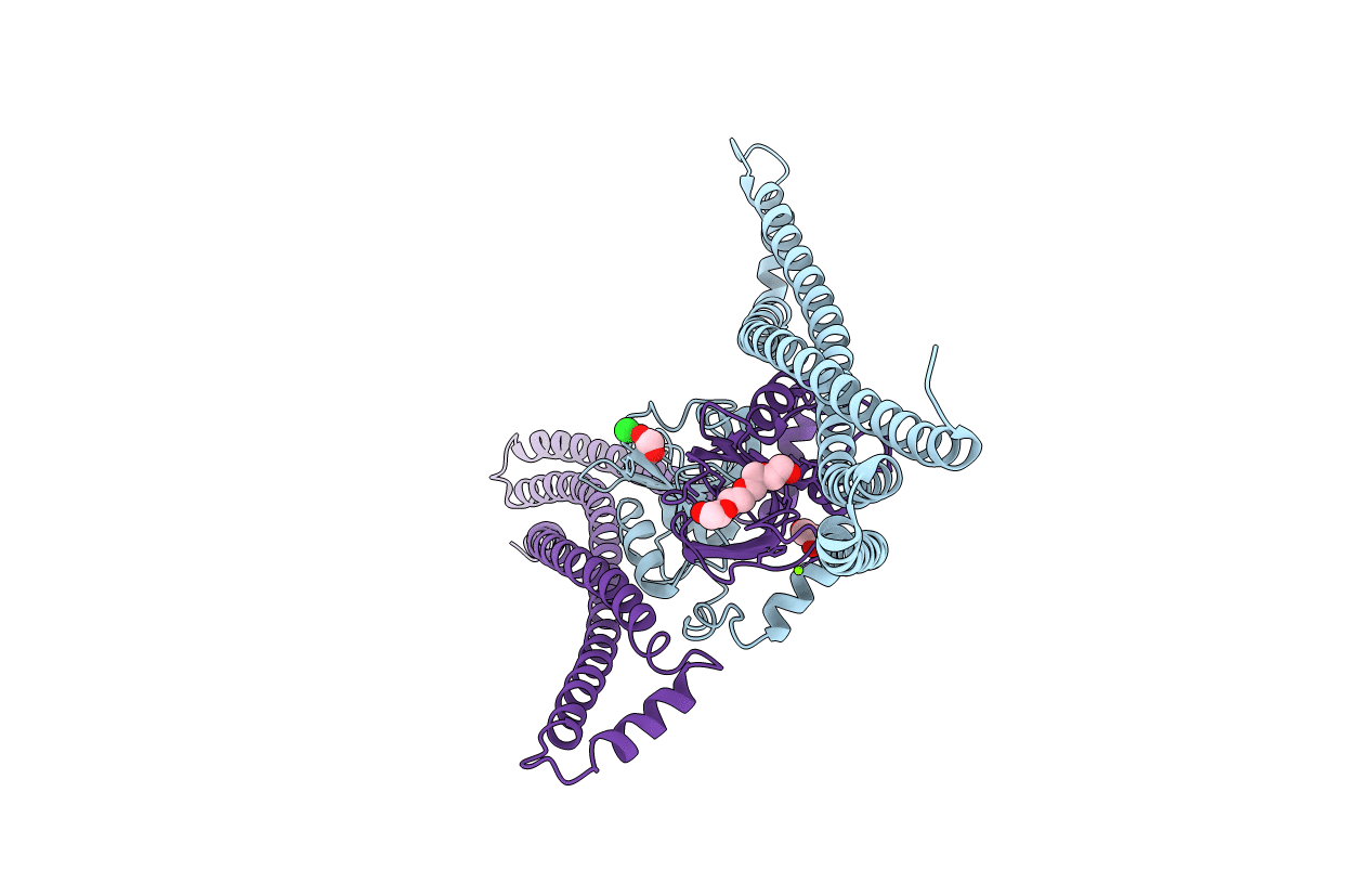
Crystal structure of the essential protein PcsB from Streptococcus pneumoniae
Biological Source:
Source Organism:
Host Organism:
Method Details:
Experimental Method:
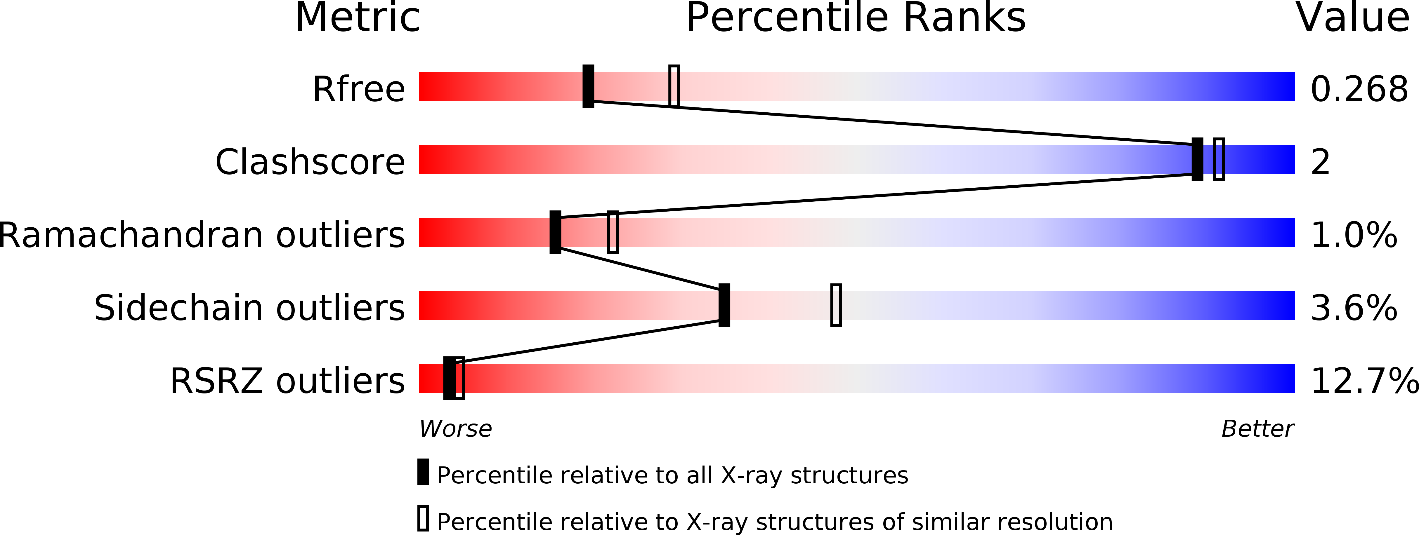
Resolution:
2.55 Å
R-Value Free:
0.27
R-Value Work:
0.23
R-Value Observed:
0.23
Space Group:
P 31 2 1
Macromolecular Entities
Polymer Type:polypeptide(L)
Molecule:SECRETED 45 KDA PROTEIN
Chain IDs:A, B
Chain Length:392
Number of Molecules:2
Biological Source:STREPTOCOCCUS PNEUMONIAE
Primary Citation
Structural Basis of Pcsb-Mediated Cell Separation in Streptococcus Pneumoniae.
Nat.Commun. 5 3842 ? (2014)
PMID: 24804636 DOI: 10.1038/NCOMMS4842

Abstact

Separation of daughter cells during bacterial cell division requires splitting of the septal cross wall by peptidoglycan hydrolases. In Streptococcus pneumoniae, PcsB is predicted to perform this operation. Recent evidence shows that PcsB is recruited to the septum by the transmembrane FtsEX complex, and that this complex is required for cell division. However, PcsB lacks detectable catalytic activity in vitro, and while it has been proposed that FtsEX activates PcsB, evidence for this is lacking. Here we demonstrate that PcsB has muralytic activity, and report the crystal structure of full-length PcsB. The protein adopts a dimeric structure in which the V-shaped coiled-coil (CC) domain of each monomer acts as a pair of molecular tweezers locking the catalytic domain of each dimeric partner in an inactive configuration. This suggests that the release of the catalytic domains likely requires an ATP-driven conformational change in the FtsEX complex, conveyed towards the catalytic domains through coordinated movements of the CC domain.

Legend

Protein

Chemical

Disease

Primary Citation of related structures