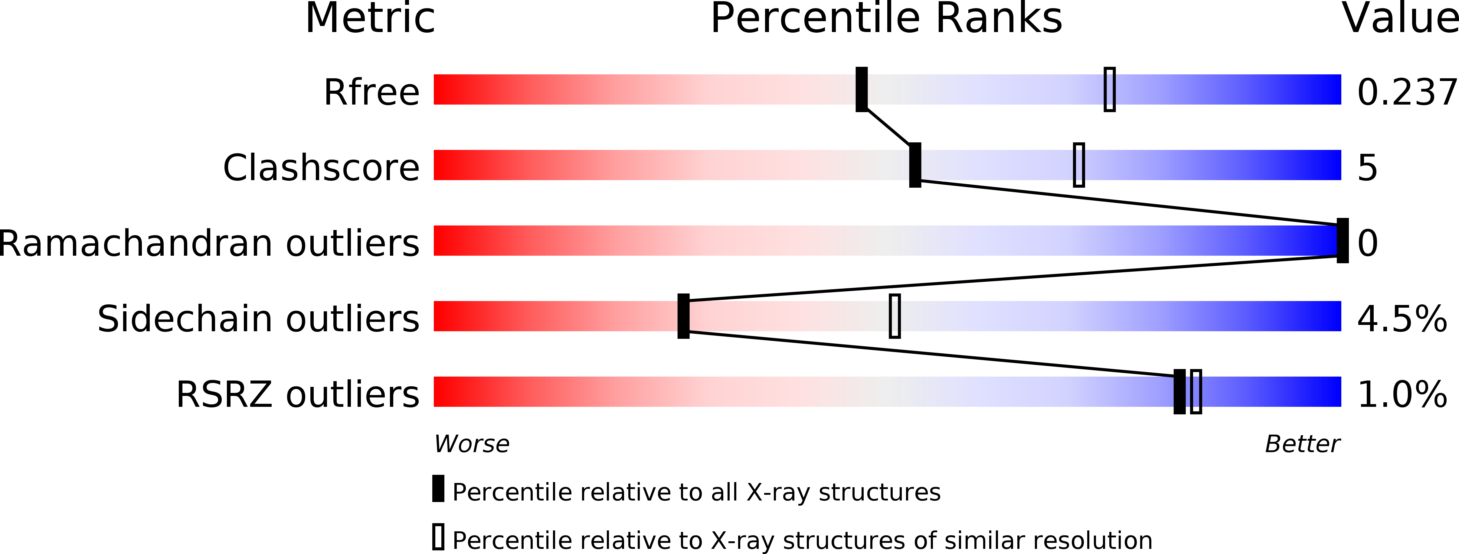
4CDO image
Deposition Date 2013-11-05
Release Date 2014-04-30
Last Version Date 2024-05-08
Entry Detail
PDB ID:
4CDO
Keywords:
Title:
Crystal structure of PQBP1 bound to spliceosomal U5-15kD
Biological Source:
Source Organism:
HOMO SAPIENS (Taxon ID: 9606)
Host Organism:
Method Details:
Experimental Method:
Resolution:
2.50 Å
R-Value Free:
0.23
R-Value Work:
0.18
R-Value Observed:
0.18
Space Group:
P 1 21 1
Macromolecular Entities
Protein Blast
Polymer Type:polypeptide(L)
Molecule:THIOREDOXIN-LIKE PROTEIN 4A, POLYGLUTAMINE-BINDING PROTEIN
Gene (Uniprot):PQBP1, TXNL4A
Chain IDs:A, B (auth: C)
Chain Length:185
Number of Molecules:2
Biological Source:HOMO SAPIENS
Primary Citation
Mutations in the Pqbp1 Gene Prevent its Interaction with the Spliceosomal Protein U5-15Kd.
Nat.Commun. 5 3822 ? (2014)
PMID: 24781215 DOI: 10.1038/NCOMMS4822

Abstact

A loss-of-function of polyglutamine tract-binding protein 1 (PQBP1) induced by frameshift mutations is believed to cause X-linked mental retardation. However, the mechanism by which structural changes in PQBP1 lead to mental retardation is unknown. Here we present the crystal structure of a C-terminal fragment of PQBP1 in complex with the spliceosomal protein U5-15 kD. The U5-15 kD hydrophobic groove recognizes a YxxPxxVL motif in PQBP1, and mutations within this motif cause a loss-of-function phenotype of PQBP1 in vitro. The YxxPxxVL motif is absent in all PQBP1 frameshift mutants seen in cases of mental retardation. These results suggest a mechanism by which the loss of the YxxPxxVL motif could lead to the functional defects seen in this type of mental retardation.

Legend

Protein

Chemical

Disease

Primary Citation of related structures
Feedback Form
Name
Email
Institute
Feedback