4CD6 image
Entry Detail
PDB ID:
4CD6
Keywords:
Title:
The structure of GH113 beta-mannanase AaManA from Alicyclobacillus acidocaldarius in complex with ManIFG
Biological Source:
Host Organism:
PDB Version:
Deposition Date:
2013-10-30
Release Date:
2014-04-02
Method Details:
Experimental Method:
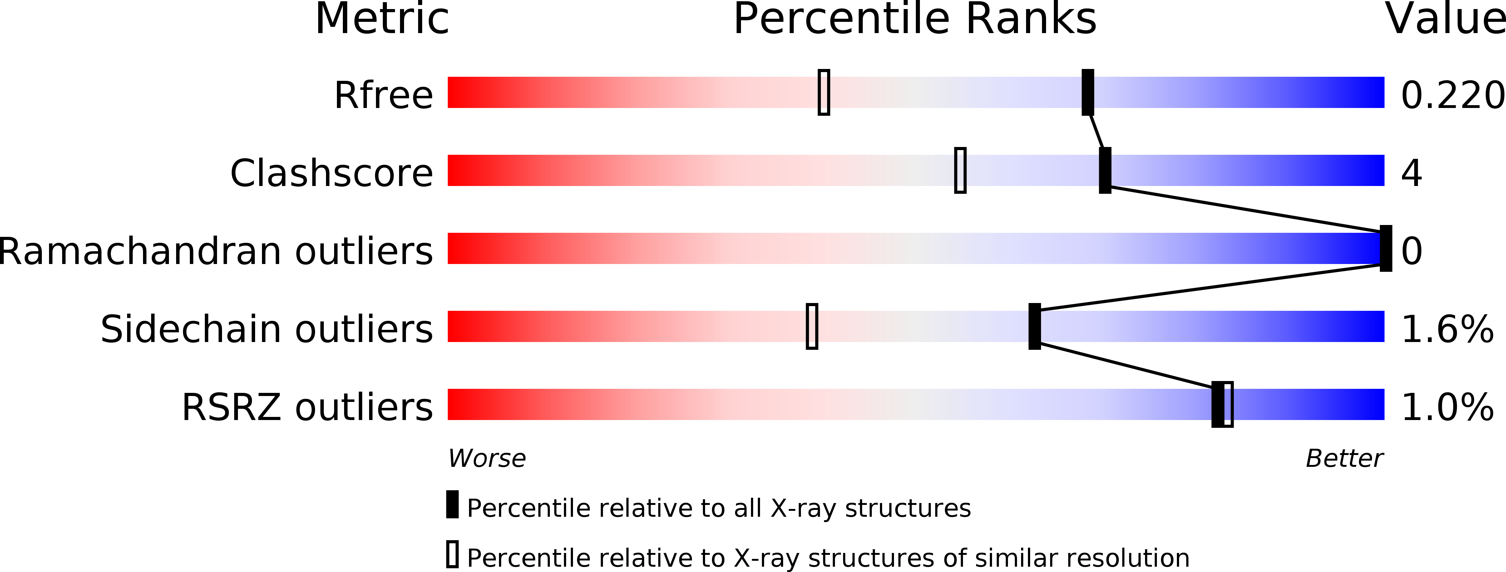
Resolution:
1.64 Å
R-Value Free:
0.20
R-Value Work:
0.14
R-Value Observed:
0.14
Space Group:
P 21 21 21
Macromolecular Entities
Polymer Type:polypeptide(L)
Description:ENDO-BETA-1,4-MANNANASE
Chain IDs:A
Chain Length:320
Number of Molecules:1
Biological Source:ALICYCLOBACILLUS ACIDOCALDARIUS
Primary Citation
Combined Inhibitor Free-Energy Landscape and Structural Analysis Reports on the Mannosidase Conformational Coordinate.
Angew.Chem.Int.Ed.Engl. 53 1087 ? (2014)
PMID: 24339341 DOI: 10.1002/ANIE.201308334

Abstact

Mannosidases catalyze the hydrolysis of a diverse range of polysaccharides and glycoconjugates, and the various sequence-based mannosidase families have evolved ingenious strategies to overcome the stereoelectronic challenges of mannoside chemistry. Using a combination of computational chemistry, inhibitor design and synthesis, and X-ray crystallography of inhibitor/enzyme complexes, it is demonstrated that mannoimidazole-type inhibitors are energetically poised to report faithfully on mannosidase transition-state conformation, and provide direct evidence for the conformational itinerary used by diverse mannosidases, including β-mannanases from families GH26 and GH113. Isofagomine-type inhibitors are poor mimics of transition-state conformation, owing to the high energy barriers that must be crossed to attain mechanistically relevant conformations, however, these sugar-shaped heterocycles allow the acquisition of ternary complexes that span the active site, thus providing valuable insight into active-site residues involved in substrate recognition.

Legend

Protein

Chemical

Disease

Primary Citation of related structures