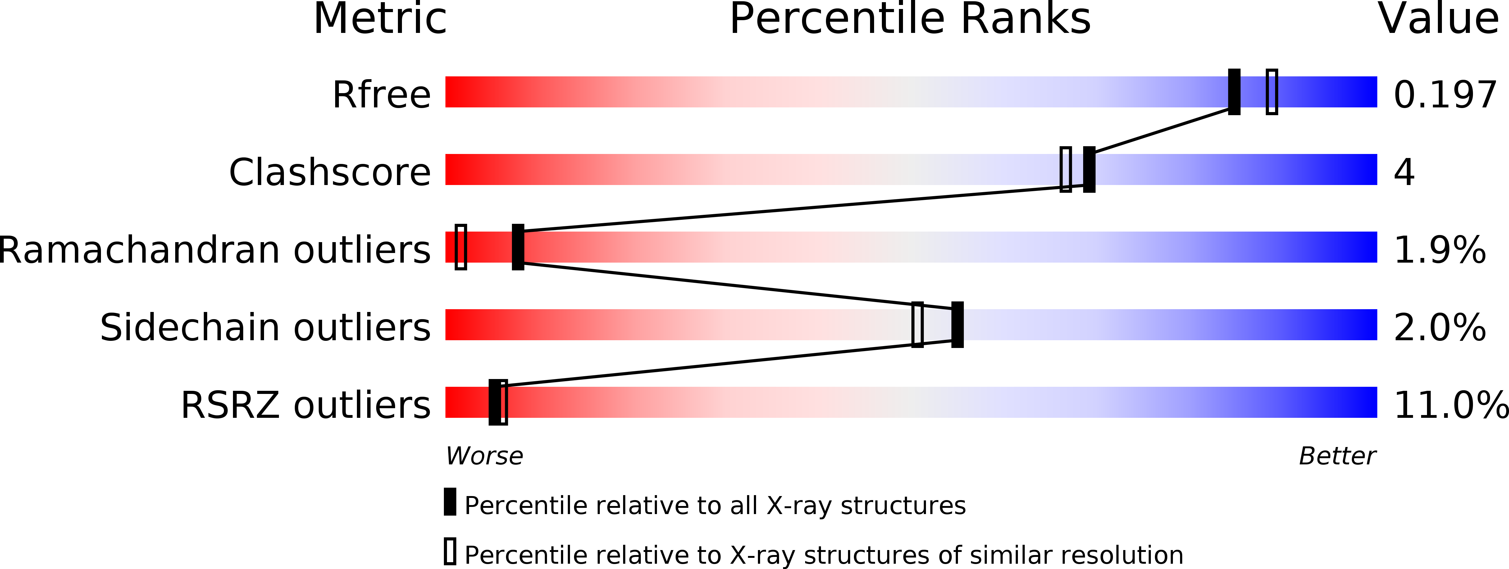
4CAH image
Deposition Date 2013-10-08
Release Date 2014-01-29
Last Version Date 2023-12-20
Entry Detail
PDB ID:
4CAH
Title:
Structure of inner DysF domain of human dysferlin
Biological Source:
Source Organism:
HOMO SAPIENS (Taxon ID: 9606)
Host Organism:
Method Details:
Experimental Method:
Resolution:
1.90 Å
R-Value Free:
0.19
R-Value Work:
0.17
R-Value Observed:
0.17
Space Group:
P 21 3
Macromolecular Entities
Polymer Type:polypeptide(L)
Molecule:DYSFERLIN
Gene (Uniprot):DYSF
Chain IDs:A (auth: B)
Chain Length:112
Number of Molecules:1
Biological Source:HOMO SAPIENS
Primary Citation
Crystal Structures of the Human Dysferlin Inner Dysf Domain
Bmc Struct.Biol. 14 3 ? (2014)
PMID: 24438169 DOI: 10.1186/1472-6807-14-3

Abstact

BACKGROUND Mutations in dysferlin, the first protein linked with the cell membrane repair mechanism, causes a group of muscular dystrophies called dysferlinopathies. Dysferlin is a type two-anchored membrane protein, with a single C terminal trans-membrane helix, and most of the protein lying in cytoplasm. Dysferlin contains several C2 domains and two DysF domains which are nested one inside the other. Many pathogenic point mutations fall in the DysF domain region. RESULTS We describe the crystal structure of the human dysferlin inner DysF domain with a resolution of 1.9 Ångstroms. Most of the pathogenic mutations are part of aromatic/arginine stacks that hold the domain in a folded conformation. The high resolution of the structure show that these interactions are a mixture of parallel ring/guanadinium stacking, perpendicular H bond stacking and aliphatic chain packing. CONCLUSIONS The high resolution structure of the Dysferlin DysF domain gives a template on which to interpret in detail the pathogenic mutations that lead to disease.

Legend

Protein

Chemical

Disease

Primary Citation of related structures