Deposition Date
2013-10-08
Release Date
2013-11-27
Last Version Date
2024-10-16
Entry Detail
PDB ID:
4CAD
Keywords:
Title:
Mechanism of farnesylated CAAX protein processing by the integral membrane protease Rce1
Biological Source:
Source Organism(s):
METHANOCOCCUS MARIPALUDIS (Taxon ID: 39152)
MUS MUSCULUS (Taxon ID: 10090)
MUS MUSCULUS (Taxon ID: 10090)
Expression System(s):
Method Details:
Experimental Method:
Resolution:
2.50 Å
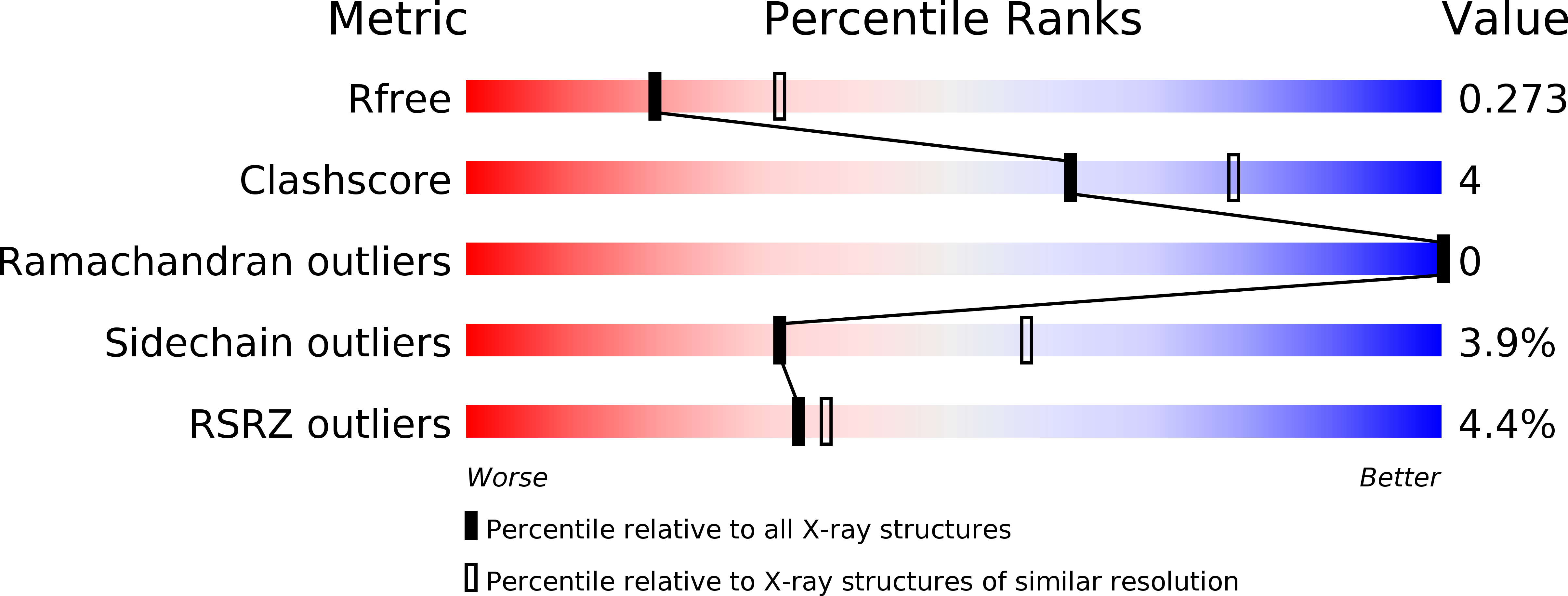
R-Value Free:
0.26
R-Value Work:
0.22
R-Value Observed:
0.22
Space Group:
P 1