4C4A image
Entry Detail
PDB ID:
4C4A
Keywords:
Title:
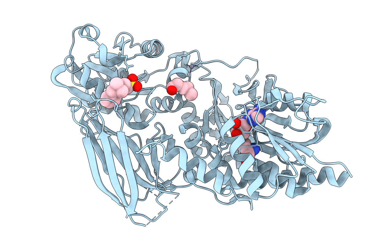
Crystal structure of mouse protein arginine methyltransferase 7 in complex with SAH
Biological Source:
Source Organism:
PDB Version:
Deposition Date:
2013-09-02
Release Date:
2014-07-02
Method Details:
Experimental Method:
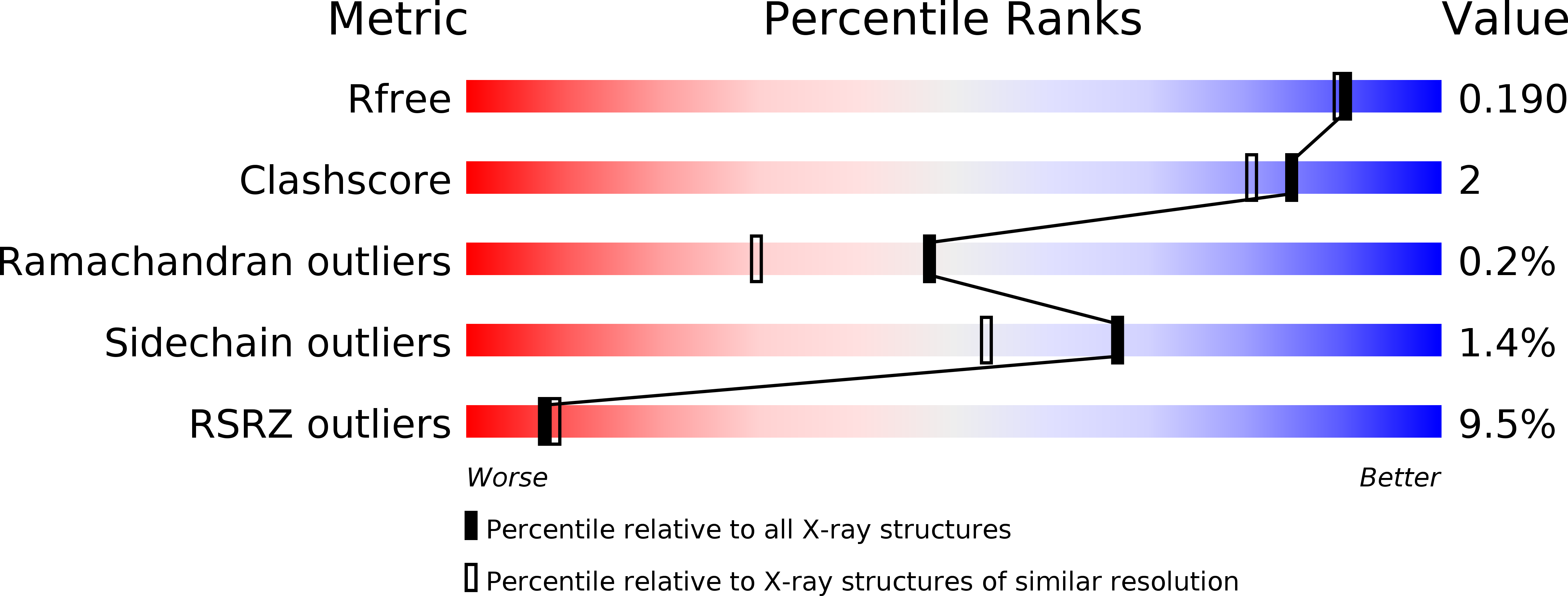
Resolution:
1.70 Å
R-Value Free:
0.18
R-Value Work:
0.15
R-Value Observed:
0.16
Space Group:
P 43 21 2
Macromolecular Entities
Polymer Type:polypeptide(L)
Description:PROTEIN ARGININE N-METHYLTRANSFERASE 7
Chain IDs:A
Chain Length:692
Number of Molecules:1
Biological Source:MUS MUSCULUS
Primary Citation
Structural Insight Into Arginine Methylation by the Mouse Protein Arginine Methyltransferase 7: A Zinc Finger Freezes the Mimic of the Dimeric State Into a Single Active Site.
Acta Crystallogr.,Sect.D 70 2401 ? (2014)
PMID: 25195753 DOI: 10.1107/S1399004714014278

Abstact

Protein arginine methyltransferase 7 (PRMT7) is a type III arginine methyltransferase which has been implicated in several biological processes such as transcriptional regulation, DNA damage repair, RNA splicing, cell differentiation and metastasis. PRMT7 is a unique but less characterized member of the family of PRMTs. The crystal structure of full-length PRMT7 from Mus musculus refined at 1.7 Å resolution is described. The PRMT7 structure is composed of two catalytic modules in tandem forming a pseudo-dimer and contains only one AdoHcy molecule bound to the N-terminal module. The high-resolution crystal structure presented here revealed several structural features showing that the second active site is frozen in an inactive state by a conserved zinc finger located at the junction between the two PRMT modules and by the collapse of two degenerated AdoMet-binding loops.

Legend

Protein

Chemical

Disease

Primary Citation of related structures