4C3X image
Deposition Date 2013-08-28
Release Date 2013-11-06
Last Version Date 2024-05-08
Entry Detail
PDB ID:
4C3X
Keywords:
Title:
Crystal structure of 3-ketosteroid delta1-dehydrogenase from Rhodococcus erythropolis SQ1
Biological Source:
Source Organism:
Host Organism:
Method Details:
Experimental Method:
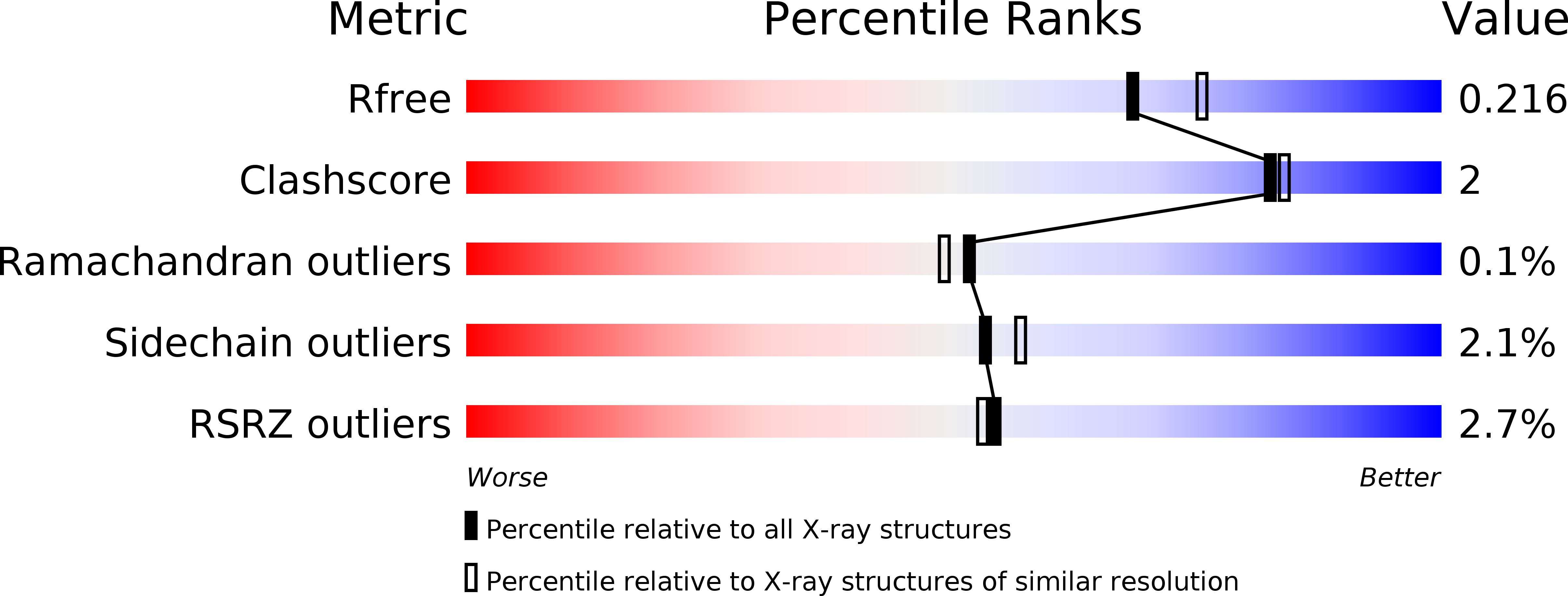
Resolution:
2.00 Å
R-Value Free:
0.20
R-Value Work:
0.17
R-Value Observed:
0.17
Space Group:
P 21 21 21
Macromolecular Entities
Structures with similar UniProt ID
Protein Blast
Polymer Type:polypeptide(L)
Molecule:3-KETOSTEROID DEHYDROGENASE
Gene (Uniprot):kstD1
Chain IDs:A, B, C, D, E, F, G, H
Chain Length:530
Number of Molecules:8
Biological Source:RHODOCOCCUS ERYTHROPOLIS
Peptide-like Molecules
PRD_900003
Primary Citation
Crystal Structure and Site-Directed Mutagenesis of 3-Ketosteroid Delta1-Dehydrogenase from Rhodococcus Erythropolis Sq1 Explain its Catalytic Mechanism
J.Biol.Chem. 288 35559 ? (2013)
PMID: 24165124 DOI: 10.1074/JBC.M113.522771

Abstact

3-Ketosteroid Δ(1)-dehydrogenases are FAD-dependent enzymes that catalyze the 1,2-desaturation of 3-ketosteroid substrates to initiate degradation of the steroid nucleus. Here we report the 2.0 Å resolution crystal structure of the 56-kDa enzyme from Rhodococcus erythropolis SQ1 (Δ(1)-KSTD1). The enzyme contains two domains: an FAD-binding domain and a catalytic domain, between which the active site is situated as evidenced by the 2.3 Å resolution structure of Δ(1)-KSTD1 in complex with the reaction product 1,4-androstadiene-3,17-dione. The active site contains four key residues: Tyr(119), Tyr(318), Tyr(487), and Gly(491). Modeling of the substrate 4-androstene-3,17-dione at the position of the product revealed its interactions with these residues and the FAD. The C1 and C2 atoms of the substrate are at reaction distance to the N5 atom of the isoalloxazine ring of FAD and the hydroxyl group of Tyr(318), respectively, whereas the C3 carbonyl group is at hydrogen bonding distance from the hydroxyl group of Tyr(487) and the backbone amide of Gly(491). Site-directed mutagenesis of the tyrosines to phenylalanines confirmed their importance for catalysis. The structural features and the kinetic properties of the mutants suggest a catalytic mechanism in which Tyr(487) and Gly(491) work in tandem to promote keto-enol tautomerization and increase the acidity of the C2 hydrogen atoms of the substrate. With assistance of Tyr(119), the general base Tyr(318) abstracts the axial β-hydrogen from C2 as a proton, whereas the FAD accepts the axial α-hydrogen from the C1 atom of the substrate as a hydride ion.

Legend

Protein

Chemical

Disease

Primary Citation of related structures