Deposition Date
2013-08-26
Release Date
2014-05-28
Last Version Date
2023-12-20
Entry Detail
PDB ID:
4C3R
Keywords:
Title:
Structure of dephosphorylated Aurora A (122-403) bound to AMPPCP
Biological Source:
Source Organism(s):
HOMO SAPIENS (Taxon ID: 9606)
Expression System(s):
Method Details:
Experimental Method:
Resolution:
2.79 Å
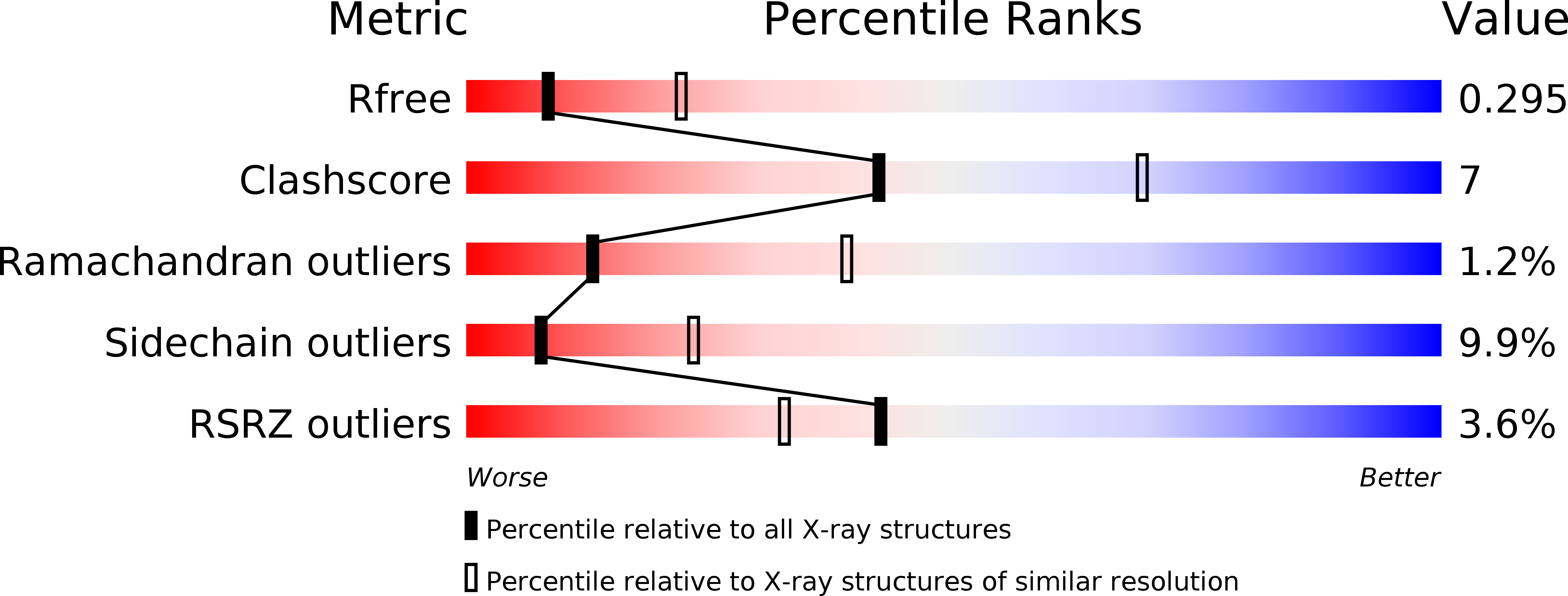
R-Value Free:
0.30
R-Value Work:
0.22
R-Value Observed:
0.22
Space Group:
P 61 2 2