4C30 image
Deposition Date 2013-08-21
Release Date 2013-10-30
Last Version Date 2024-05-01
Entry Detail
PDB ID:
4C30
Keywords:
Title:
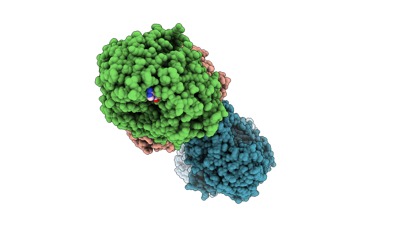
Crystal structure of Deinococcus radiodurans UvrD in complex with DNA, form 2
Biological Source:
Source Organism:
Host Organism:
Method Details:
Experimental Method:
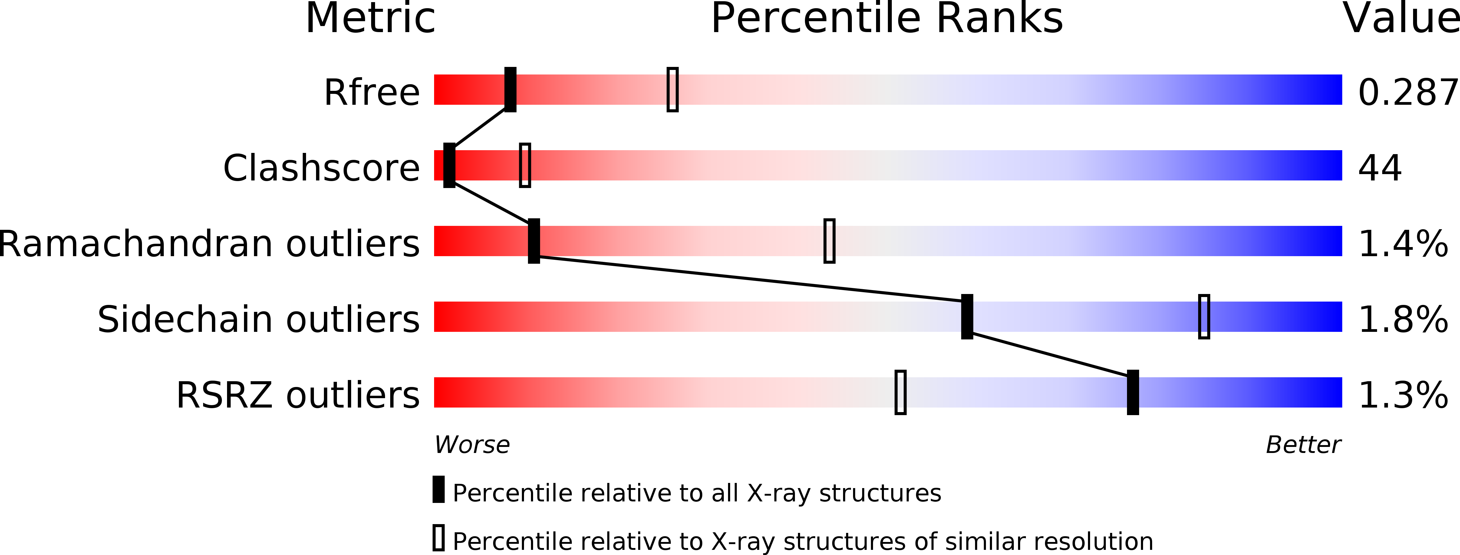
Resolution:
3.00 Å
R-Value Free:
0.28
R-Value Work:
0.22
R-Value Observed:
0.23
Space Group:
P 1 21 1
Macromolecular Entities
Polymer Type:polypeptide(L)
Molecule:DNA HELICASE II
Gene (Uniprot):DR_1775
Chain IDs:A, B (auth: D), C (auth: F), D (auth: I)
Chain Length:665
Number of Molecules:4
Biological Source:DEINOCOCCUS RADIODURANS
Polymer Type:polydeoxyribonucleotide
Molecule:DNA STRAND FOR25
Chain IDs:E (auth: K), G (auth: X)
Chain Length:25
Number of Molecules:2
Biological Source:SYNTHETIC CONSTRUCT
Polymer Type:polydeoxyribonucleotide
Molecule:DNA STRAND REV25
Chain IDs:F (auth: L), H (auth: Y)
Chain Length:25
Number of Molecules:2
Biological Source:SYNTHETIC CONSTRUCT
Primary Citation
Structural and Mechanistic Insight Into DNA Unwinding by Deinococcus Radiodurans Uvrd.
Plos One 8 77364 ? (2013)
PMID: 24143224 DOI: 10.1371/JOURNAL.PONE.0077364

Abstact

DNA helicases are responsible for unwinding the duplex DNA, a key step in many biological processes. UvrD is a DNA helicase involved in several DNA repair pathways. We report here crystal structures of Deinococcus radiodurans UvrD (drUvrD) in complex with DNA in different nucleotide-free and bound states. These structures provide us with three distinct snapshots of drUvrD in action and for the first time trap a DNA helicase undergoing a large-scale spiral movement around duplexed DNA. Our structural data also improve our understanding of the molecular mechanisms that regulate DNA unwinding by Superfamily 1A (SF1A) helicases. Our biochemical data reveal that drUvrD is a DNA-stimulated ATPase, can translocate along ssDNA in the 3'-5' direction and shows ATP-dependent 3'-5', and surprisingly also, 5'-3' helicase activity. Interestingly, we find that these translocase and helicase activities of drUvrD are modulated by the ssDNA binding protein. Analysis of drUvrD mutants indicate that the conserved β-hairpin structure of drUvrD that functions as a separation pin is critical for both drUvrD's 3'-5' and 5'-3' helicase activities, whereas the GIG motif of drUvrD involved in binding to the DNA duplex is essential for the 5'-3' helicase activity only. These special features of drUvrD may reflect its involvement in a wide range of DNA repair processes in vivo.

Legend

Protein

Chemical

Disease

Primary Citation of related structures