4C2L image
Deposition Date 2013-08-19
Release Date 2013-09-25
Last Version Date 2024-10-23
Entry Detail
PDB ID:
4C2L
Keywords:
Title:
Crystal structure of endo-xylogalacturonan hydrolase from Aspergillus tubingensis
Biological Source:
Source Organism:
Host Organism:
Method Details:
Experimental Method:
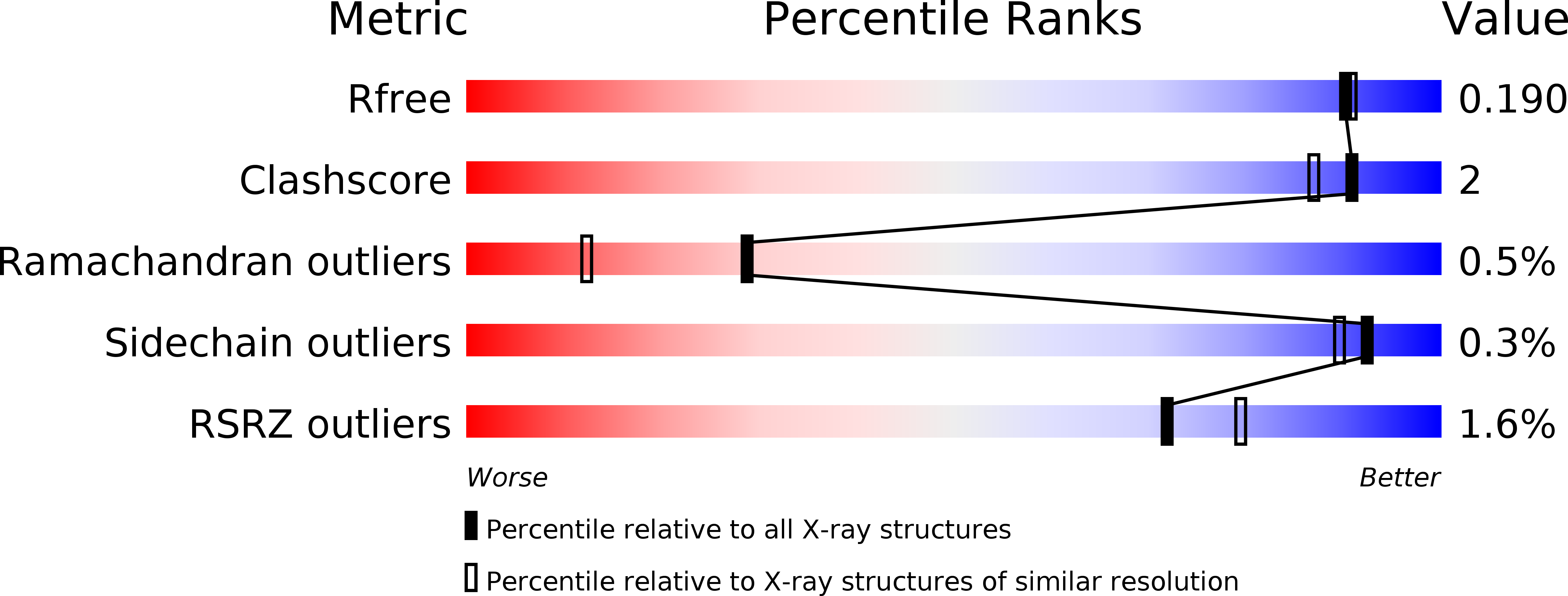
Resolution:
1.75 Å
R-Value Free:
0.18
R-Value Work:
0.16
R-Value Observed:
0.16
Space Group:
I 2 2 2
Macromolecular Entities
Structures with similar UniProt ID
Protein Blast
Polymer Type:polypeptide(L)
Molecule:ENDO-XYLOGALACTURONAN HYDROLASE A
Gene (Uniprot):xghA
Chain IDs:A
Chain Length:388
Number of Molecules:1
Biological Source:ASPERGILLUS TUBINGENSIS
Modified Residue
Compound ID Chain ID Parent Comp ID Details 2D Image
ASN A ASN GLYCOSYLATION SITE
THR A THR GLYCOSYLATION SITE
Primary Citation
Crystal Structure of Endo-Xylogalacturonan Hydrolase from Aspergillus Tubingensis.
FEBS J. 280 6061 ? (2013)
PMID: 24034788 DOI: 10.1111/FEBS.12524

Abstact

UNLABELLED Endo-xylogalacturonan hydrolase is a member of glycoside hydrolase family 28 (GH28) that hydrolyzes the glycosidic bond between two β-xylose-substituted galacturonic acid residues in pectin. Presented here is the X-ray crystal structure of the endo-xylogalacturonan hydrolase from Aspergillus tubingensis (XghA) at 1.75 Å resolution. The high degree of structural conservation in the active site and catalytic apparatus compared with polygalacturonases indicates that cleavage of the substrate proceeds in essentially the same way as found for the other GH28 enzymes. Molecular modeling of a xylosylated tri-galacturonate in the active site identified the amino acid residues involved in substrate binding. They border a substrate-binding cleft that is much wider than in other polygalacturonases, and can accommodate xylosylated substrates. The most extensive interactions appear to occur at subsite +2, in agreement with the enzyme kinetics results, which showed enhanced activity on substrates with a xylose attached to the galacturonic acid bound at subsite +2. DATABASE Structural data are available in the Protein Data Bank database under accession number 4C2L.

Legend

Protein

Chemical

Disease

Primary Citation of related structures
Feedback Form
Name
Email
Institute
Feedback