4C1X image
Deposition Date 2013-08-14
Release Date 2014-09-10
Last Version Date 2024-05-08
Entry Detail
PDB ID:
4C1X
Keywords:
Title:
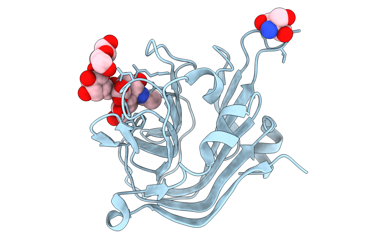
Carbohydrate binding domain from Streptococcus pneumoniae NanA sialidase complexed with 6'-sialyllactose
Biological Source:
Source Organism:
Host Organism:
Method Details:
Experimental Method:
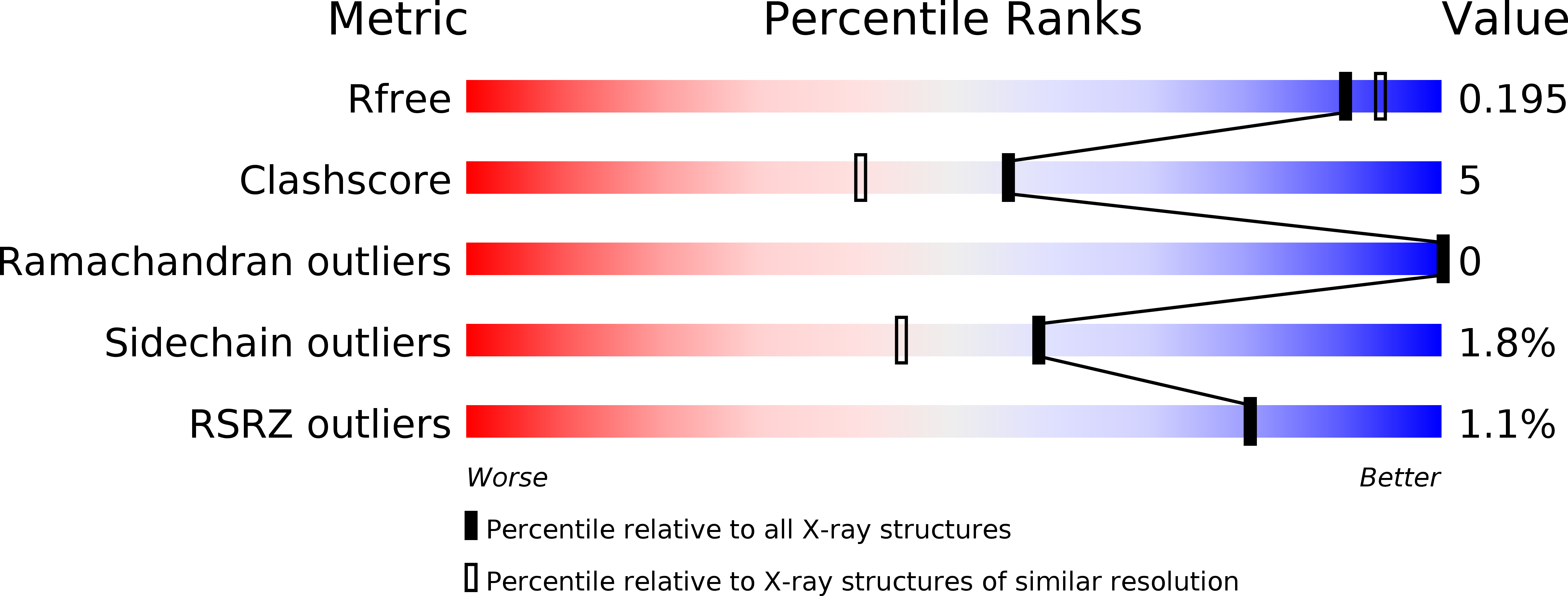
Resolution:
1.84 Å
R-Value Free:
0.19
R-Value Work:
0.14
R-Value Observed:
0.15
Space Group:
P 1 21 1
Macromolecular Entities
Structures with similar UniProt ID
Protein Blast
Polymer Type:polypeptide(L)
Molecule:SIALIDASE A
Gene (Uniprot):nanA
Chain IDs:A
Chain Length:188
Number of Molecules:1
Biological Source:STREPTOCOCCUS PNEUMONIAE
Ligand Molecules
Peptide-like Molecules
PRD_900066
Primary Citation
Structural Characterization of the Carbohydrate-Binding Module of Nana Sialidase, a Pneumococcal Virulence Factor.
Bmc Struct.Biol. 15 15 ? (2015)
PMID: 26289431 DOI: 10.1186/S12900-015-0042-4

Abstact

BACKGROUND Streptococcus pneumoniae Neuraminidase A (NanA) is a multi-domain protein anchored to the bacterial surface. Upstream of the catalytic domain of NanA is a domain that conforms to the sialic acid-recognising CBM40 family of the CAZY (carbohydrate-active enzymes) database. This domain has been identified to play a critical role in allowing the bacterium to promote adhesion and invasion of human brain microvascular endothelial cells, and hence may play a key role in promoting bacterial meningitis. In addition, the CBM40 domain has also been reported to activate host chemokines and neutrophil recruitment during infection. RESULTS Crystal structures of both apo- and holo- forms of the NanA CBM40 domain (residues 121 to 305), have been determined to 1.8 Å resolution. The domain shares the fold of other CBM40 domains that are associated with sialidases. When in complex with α2,3- or α2,6-sialyllactose, the domain is shown to interact only with the terminal sialic acid. Significantly, a deep acidic pocket adjacent to the sialic acid-binding site is identified, which is occupied by a lysine from a symmetry-related molecule in the crystal. This pocket is adjacent to a region that is predicted to be involved in protein-protein interactions. CONCLUSIONS The structural data provide the details of linkage-independent sialyllactose binding by NanA CBM40 and reveal striking surface features that may hold the key to recognition of binding partners on the host cell surface. The structure also suggests that small molecules or sialic acid analogues could be developed to fill the acidic pocket and hence provide a new therapeutic avenue against meningitis caused by S. pneumoniae.

Legend

Protein

Chemical

Disease

Primary Citation of related structures
Feedback Form
Name
Email
Institute
Feedback