4C14 image
Deposition Date 2013-08-09
Release Date 2013-10-30
Last Version Date 2023-12-20
Entry Detail
PDB ID:
4C14
Keywords:
Title:
The crystal strucuture of PpAzoR in complex with reactive black 5 (RB5)
Biological Source:
Source Organism:
Host Organism:
Method Details:
Experimental Method:
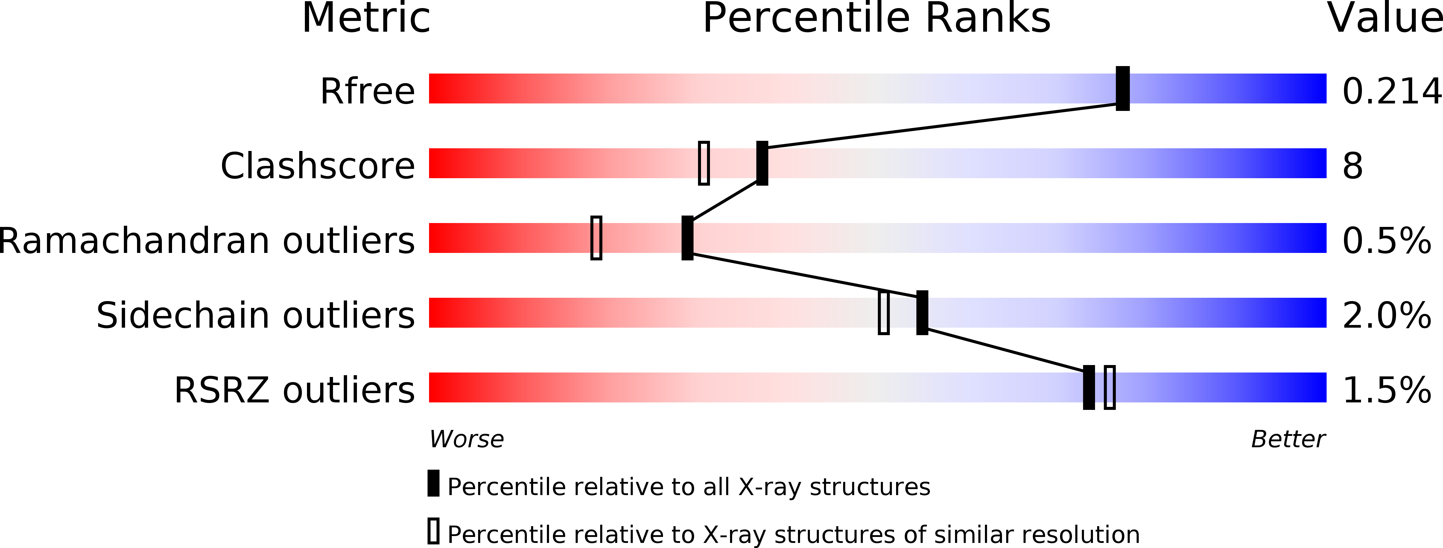
Resolution:
1.90 Å
R-Value Free:
0.21
R-Value Work:
0.18
R-Value Observed:
0.18
Space Group:
F 2 2 2
Macromolecular Entities
Polymer Type:polypeptide(L)
Molecule:FMN-DEPENDENT NADH-AZOREDUCTASE 1
Gene (Uniprot):azoR1
Chain IDs:A
Chain Length:203
Number of Molecules:1
Biological Source:PSEUDOMONAS PUTIDA
Primary Citation
The crystal structure of Pseudomonas putida azoreductase - the active site revisited.
FEBS J. 280 6643 6657 (2013)
PMID: 24127652 DOI: 10.1111/febs.12568

Abstact

The enzymatic degradation of azo dyes begins with the reduction of the azo bond. In this article, we report the crystal structures of the native azoreductase from Pseudomonas putida MET94 (PpAzoR) (1.60 Å), of PpAzoR in complex with anthraquinone-2-sulfonate (1.50 Å), and of PpAzoR in complex with Reactive Black 5 dye (1.90 Å). These structures reveal the residues and subtle changes that accompany substrate binding and release. Such changes highlight the fine control of access to the catalytic site that is required by the ping-pong mechanism, and in turn the specificity offered by the enzyme towards different substrates. The topology surrounding the active site shows novel features of substrate recognition and binding that help to explain and differentiate the substrate specificity observed among different bacterial azoreductases.

Legend

Protein

Chemical

Disease

Primary Citation of related structures