Deposition Date
2013-07-15
Release Date
2013-07-31
Last Version Date
2024-10-09
Entry Detail
PDB ID:
4BXS
Keywords:
Title:
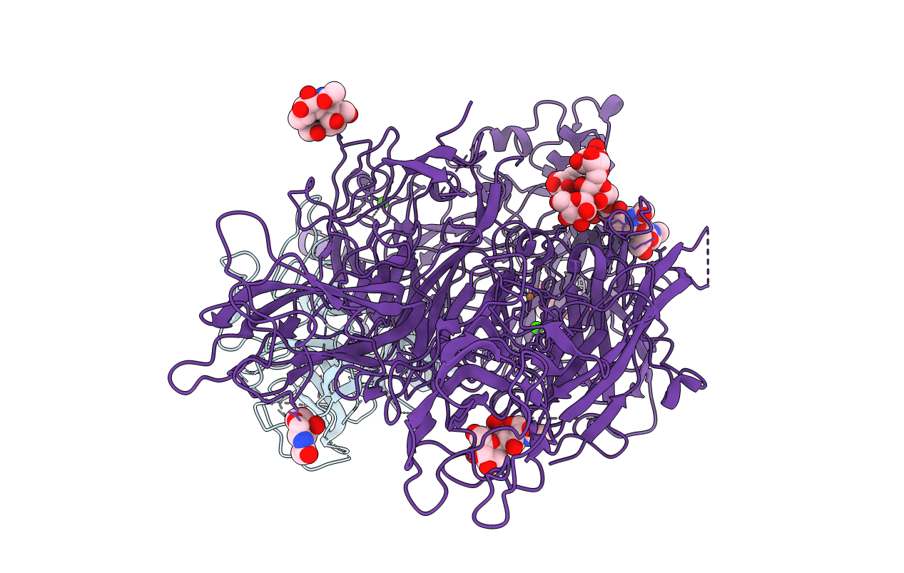
Crystal Structure of the Prothrombinase Complex from the Venom of Pseudonaja Textilis
Biological Source:
Source Organism(s):
PSEUDONAJA TEXTILIS (Taxon ID: 8673)
Expression System(s):
Method Details:
Experimental Method:
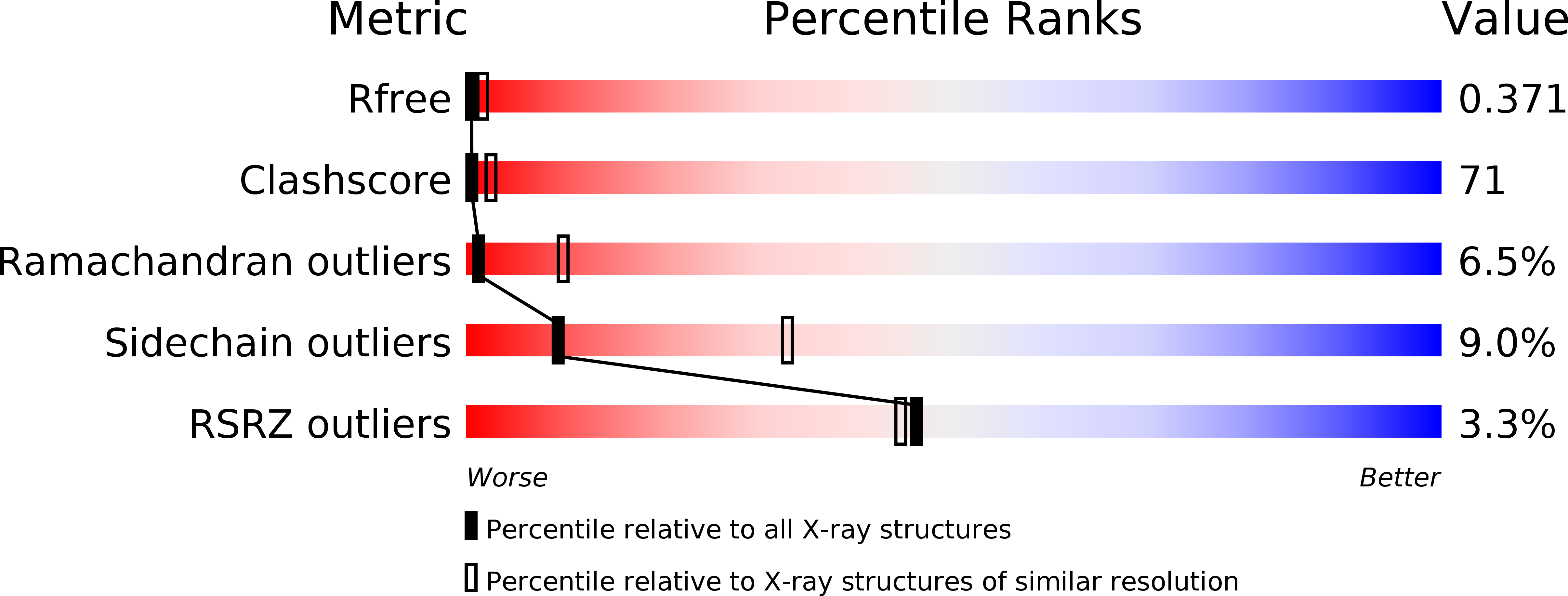
Resolution:
3.32 Å
R-Value Free:
0.36
R-Value Work:
0.29
R-Value Observed:
0.30
Space Group:
P 43 21 2