Deposition Date
2013-07-08
Release Date
2013-09-25
Last Version Date
2023-12-20
Entry Detail
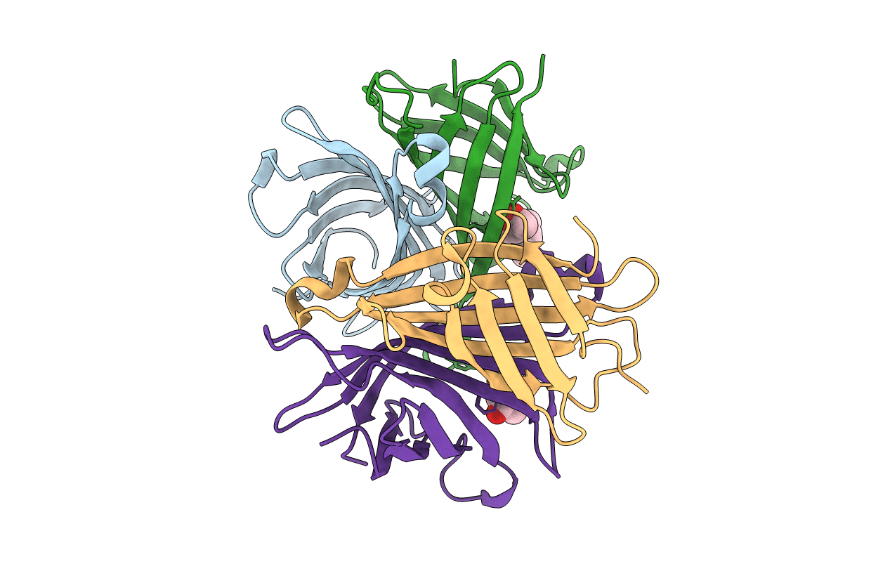
Biological Source:
Source Organism(s):
STREPTOMYCES AVIDINII (Taxon ID: 1895)
Expression System(s):
Method Details:
Experimental Method:
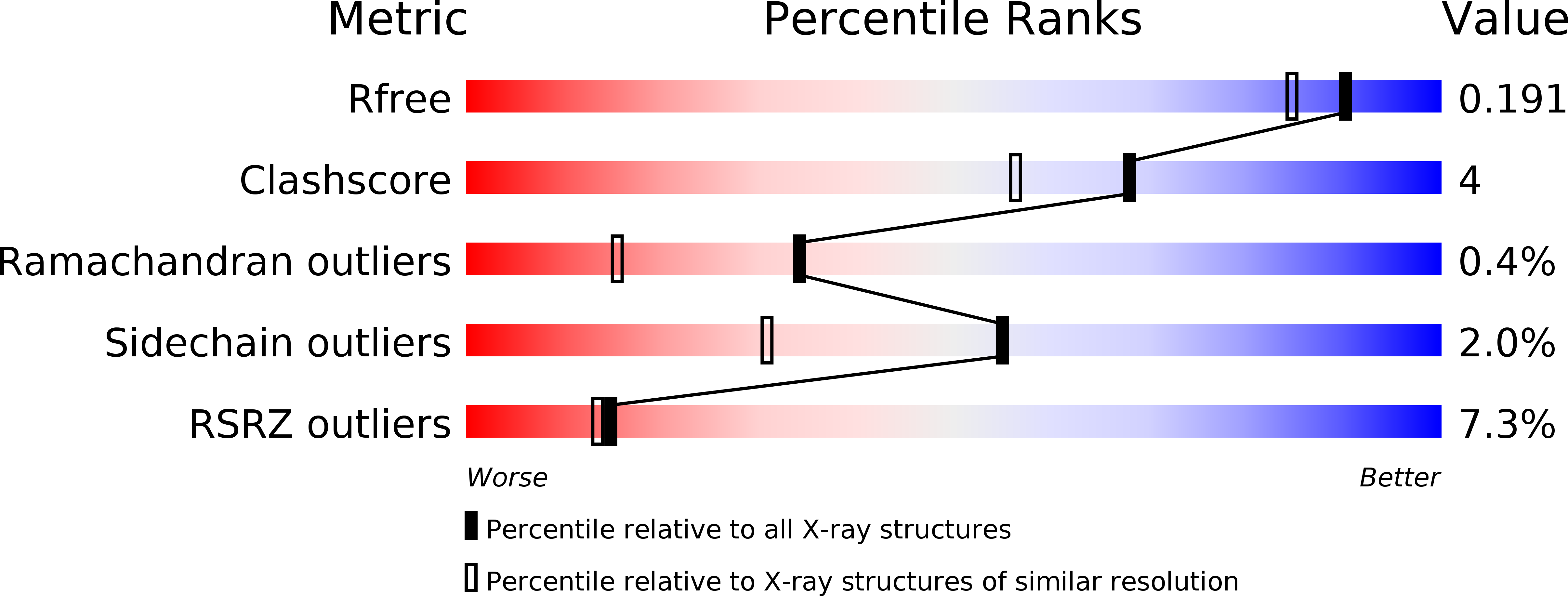
Resolution:
1.60 Å
R-Value Free:
0.18
R-Value Work:
0.16
R-Value Observed:
0.16
Space Group:
P 1 21 1