Deposition Date
2013-06-18
Release Date
2013-08-07
Last Version Date
2024-05-08
Entry Detail
PDB ID:
4BTP
Keywords:
Title:
Structure of the capsid protein P1 of the bacteriophage phi8
Biological Source:
Source Organism(s):
Pseudomonas phage phi8 (Taxon ID: 120086)
Expression System(s):
Method Details:
Experimental Method:
Resolution:
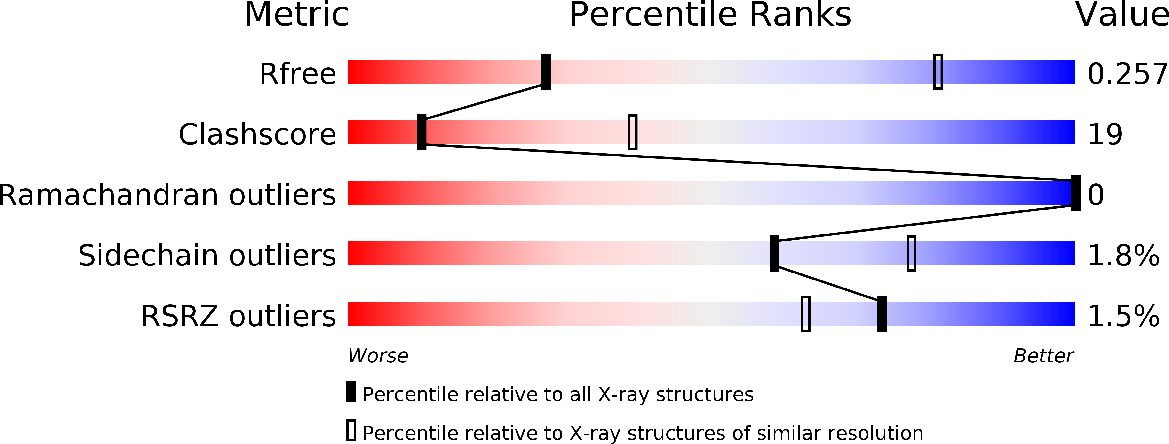
3.70 Å
R-Value Free:
0.25
R-Value Work:
0.24
R-Value Observed:
0.24
Space Group:
P 41 21 2