4BT8 image
Deposition Date 2013-06-14
Release Date 2013-10-09
Last Version Date 2023-12-20
Entry Detail
PDB ID:
4BT8
Keywords:
Title:
CRYSTAL STRUCTURE OF THE APO FORM OF N-TERMINAL DOMAIN AND PEPTIDE SUBSTRATE BINDING DOMAIN OF PROLYL-4 HYDROXYLASE TYPE I FROM HUMAN
Biological Source:
Source Organism:
HOMO SAPIENS (Taxon ID: 9606)
Host Organism:
Method Details:
Experimental Method:
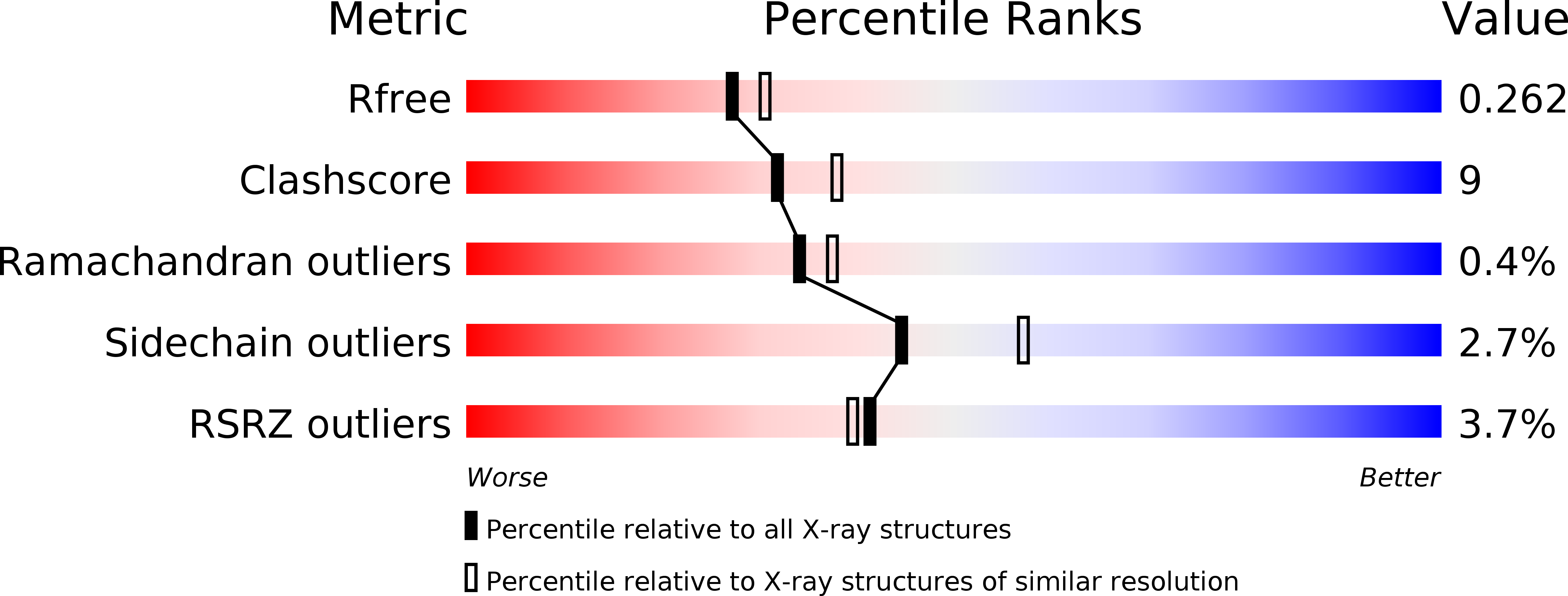
Resolution:
2.20 Å
R-Value Free:
0.26
R-Value Work:
0.21
R-Value Observed:
0.22
Space Group:
P 21 21 2
Macromolecular Entities
Polymer Type:polypeptide(L)
Molecule:PROLYL 4-HYDROXYLASE SUBUNIT ALPHA-1
Gene (Uniprot):P4HA1
Chain IDs:A, B
Chain Length:239
Number of Molecules:2
Biological Source:HOMO SAPIENS
Primary Citation
The Structural Motifs for Substrate Binding and Dimerization of the Alpha Subunit of Collagen Prolyl 4-Hydroxylase
Structure 21 2107 ? (2013)
PMID: 24207127 DOI: 10.1016/J.STR.2013.09.005

Abstact

Collagen prolyl 4-hydroxylase (C-P4H) catalyzes the proline hydroxylation of procollagen, an essential modification in the maturation of collagens. C-P4H consists of two catalytic α subunits and two protein disulfide isomerase β subunits. The assembly of these subunits is unknown. The α subunit contains an N domain (1-143), a peptide-substrate-binding-domain (PSB, 144-244) and a catalytic domain (245-517). Here, we report the dimeric structure of the N-terminal region (1-244) of the α subunit. It is shown that the N domain has an important role in the assembly of the C-P4H tetramer, by forming an extended four-helix bundle that includes an antiparallel coiled-coil dimerization motif between the two α subunits. Complexes of this construct with a C-P4H inhibitor and substrate show the mode of peptide-binding to the PSB domain. Both peptides adopt a poly-(L)-proline-type-II helix conformation and bind in a curved, asymmetric groove lined by conserved tyrosines and an Arg-Asp salt bridge.

Legend

Protein

Chemical

Disease

Primary Citation of related structures