4BRW image
Deposition Date 2013-06-05
Release Date 2013-07-24
Last Version Date 2023-12-20
Entry Detail
PDB ID:
4BRW
Keywords:
Title:
Crystal structure of the yeast Dhh1-Pat1 complex
Biological Source:
Source Organism:
Host Organism:
Method Details:
Experimental Method:
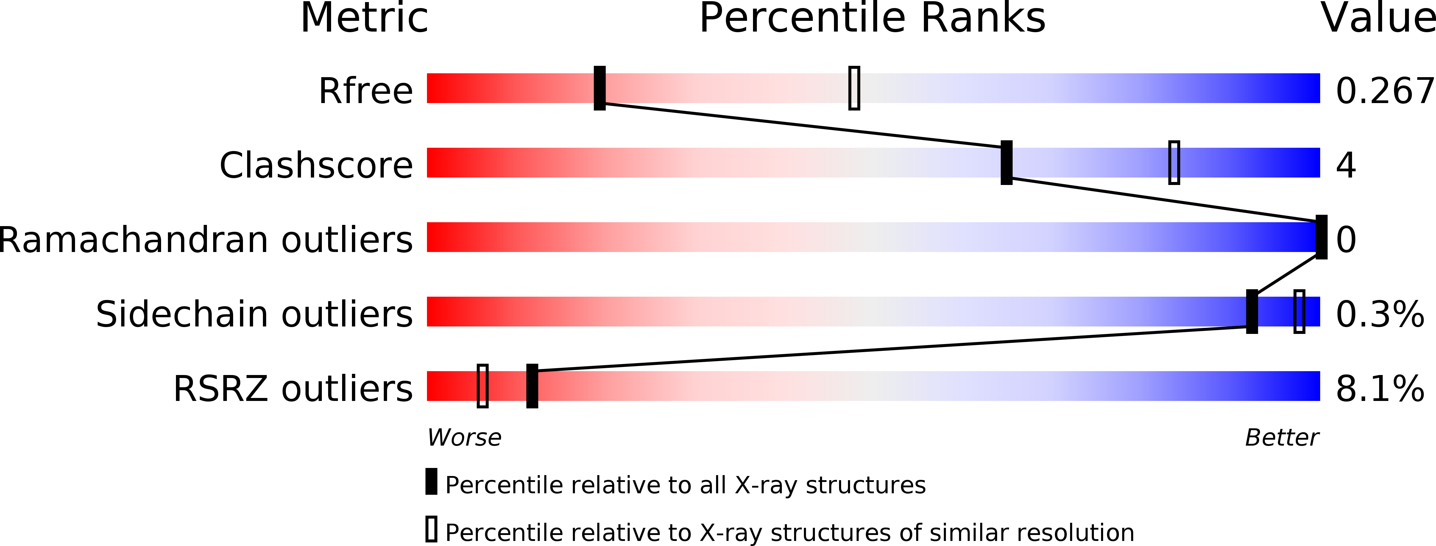
Resolution:
2.80 Å
R-Value Free:
0.24
R-Value Work:
0.20
R-Value Observed:
0.20
Space Group:
P 41 2 2
Macromolecular Entities
Structures with similar UniProt ID
Protein Blast
Polymer Type:polypeptide(L)
Molecule:ATP-DEPENDENT RNA HELICASE DHH1
Gene (Uniprot):DHH1
Mutagens:YES
Chain IDs:A
Chain Length:377
Number of Molecules:1
Biological Source:SACCHAROMYCES CEREVISIAE
Structures with similar UniProt ID
Protein Blast
Polymer Type:polypeptide(L)
Molecule:DNA TOPOISOMERASE 2-ASSOCIATED PROTEIN PAT1
Gene (Uniprot):PAT1
Chain IDs:B
Chain Length:75
Number of Molecules:1
Biological Source:SACCHAROMYCES CEREVISIAE
Primary Citation
Structural Analysis of the Yeast Dhh1-Pat1 Complex Reveals How Dhh1 Engages Pat1, Edc3 and RNA in Mutually Exclusive Interactions
Nucleic Acids Res. 41 8377 ? (2013)
PMID: 23851565 DOI: 10.1093/NAR/GKT600

Abstact

Translational repression and deadenylation of eukaryotic mRNAs result either in the sequestration of the transcripts in a nontranslatable pool or in their degradation. Removal of the 5' cap structure is a crucial step that commits deadenylated mRNAs to 5'-to-3' degradation. Pat1, Edc3 and the DEAD-box protein Dhh1 are evolutionary conserved factors known to participate in both translational repression and decapping, but their interplay is currently unclear. We report the 2.8 Å resolution structure of yeast Dhh1 bound to the N-terminal domain of Pat1. The structure shows how Pat1 wraps around the C-terminal RecA domain of Dhh1, docking onto the Phe-Asp-Phe (FDF) binding site. The FDF-binding site of Dhh1 also recognizes Edc3, revealing why the binding of Pat1 and Edc3 on Dhh1 are mutually exclusive events. Using co-immunoprecipitation assays and structure-based mutants, we demonstrate that the mode of Dhh1-Pat1 recognition is conserved in humans. Pat1 and Edc3 also interfere and compete with the RNA-binding properties of Dhh1. Mapping the RNA-binding sites on Dhh1 with a crosslinking-mass spectrometry approach shows a large RNA-binding surface around the C-terminal RecA domain, including the FDF-binding pocket. The results suggest a model for how Dhh1-containing messenger ribonucleoprotein particles might be remodeled upon Pat1 and Edc3 binding.

Legend

Protein

Chemical

Disease

Primary Citation of related structures